Surgical Approach
Preoperative mammography is essential prior to treatment of any patient with suspected malignancy. The mammogram may show the extent of the lesion, or additional lesions or microcalcification elsewhere in the breast that alter the operative approach. It may also show lesions in the other breast.
Preoperative cytology or histology must also be reviewed.
Technique of Breast Biopsy/Partial Mastectomy
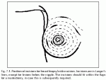
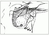
The incision for breast biopsy or wide excision is placed to allow an adequate biopsy with a good cosmetic result, which does not compromise uture treatment (i.e., it fits within usual mastectomy flaps). Extensive tunneling is avoided. For central lesions, a periareolar incision is very satisfactory, but for peripheral lesions, the incision is placed close to the lesion in Langer’s lines.
In the inferior part of the breast, a radial incision has been advocated (Fig. 7.1), although we have had better cosmetic success with the curved skin-line incision used elsewhere in the breast. To avoid tunneling when needle localization is used the incision is placed over the lesion, rather than close to the site of needle entry. If axillary dissection is required, a separate incision is used unless the lesion is located in the axillary tail.
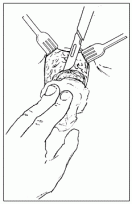
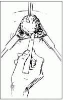
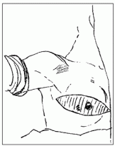
Local anaesthetic is infiltrated prior to making the incision, and the skin and subcutaneous tissue are retracted (Fig. 7.2). The breast tissue overlying the mass is grasped with Allis clamps, and the mass is excised with sharp dissection (Fig. 7.3). Electrocautery is not used for the excision, as it makes pathologic assessment of the margins more difficult. The mass itself is not grasped with clamps, as this risks tumor disruption and dissemination.
The amount of tissue removed depends on the indication. A fibroadenoma is removed with little if any surrounding breast tissue. For a small suspicious lesion, it is wise to take a margin of about 1 cm so that the diagnostic excisional biopsy might also be adequate local surgery.
Diagnostic biopsy of an equivocal area should remove the lump if it is discrete, or consist of a representative sample if it is less well defined. For a breast cancer diagnosed on percutaneous biopsy, a 1cm margin of grossly normal breast tissue is removed. An ellipse of skin can be included with a superficial tumor.
The specimen should be oriented with clips or sutures to allow the pathologist to assess the margins. After needle-localized biopsy, specimen radiography is essential to ensure complete removal of the mammographic abnormality. If cancer is suspected, it is valuable to place titanium clips in the tumor bed to acilitate radiation targeting. In cases of inflammatory or locally advanced cancer, incisional biopsy may be needed for diagnosis and treatment planning.
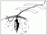
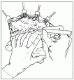
Meticulous hemostasis is obtained with electrocautery and interrupted absorbable sutures if needed. Deep breast tissue is not usually reapproximated, as this leads to later distortion. Interrupted sutures to the superficial breast tissue or subcutaneous tissue followed by a subcuticular skin suture produce an optimal cosmetic result (Fig. 7.4). Breast support is needed for at least 48 hours postoperatively.
Patrick I. Borgen and Bruce Mann
Breast conservation therapy for invasive carcinoma of the breast. Current Problems in Surgery 1995; 33:189-256.
Fig. 7.2. The skin and subcutaneous tissue is incised down to the vicinity of the mass.
Fig. 7.3. The mass is grasped with a clamp and excised using sharp dissection.
Fig. 7.4. The wound is closed with a subcuticular suture and steri-strips. The cavity is not closed.
Fig. 7.5. Skin flaps are marked to include the biopsy site. Enough skin is removed to result in a flat scar.
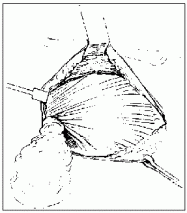
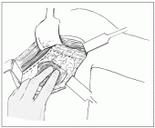
Fig. 7.6. Skin flaps are developed with upward traction on the skin, and countertraction on the breast. The breast is removed with its fascial envelope.
Fig. 7.7. The breast is removed along with the pectoralis fascia in a superomedial to inferolateral direction.
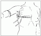
Fig. 7.8. The wound is closed over a suction drain (illustration shows a second drain in the axilla) using a subcuticular suture and steri-strips.
Fig. 7.9. Schematic diagram showing the incision for an axillary dissection and the extent to which flaps are elevated.
Fig. 7.10. Incision of the clavipectoral fascia along the lateral edge of the pectoralis minor and below the axillary vein. The position of the vein is outlined.
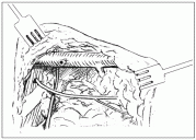
Fig. 7.11. Axillary dissection in progress. The anterior tributary of the axillary vein has been divided, the intercostobrachial nerve has been preserved and the long thoracic nerve identified.
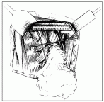
Fig. 7.12. Advanced stage of axillary dissection. The thoracodorsal nerve has been preserved, and the tissue between the long thoracic and thoracodorsal nerves has been removed.
References
- Veronesi U, Saccozzi R, Del Vecchio M et al. Comparing radical mastectomy with quadrantectomy, axillary dissection, and radiation therapy in patients with small cancers of the breast. N Engl J Med 1981; 305:6-11.
This is the landmark report of the first randomized prospective trial of breast-conservation therapy. - Fisher B, Bauer M, Margolese R et al. Five-year results of a randomized clinical trial comparing total mastectomy and segmental mastectomy with or without radiation in the treatment of breast cancer. N Engl J Med 1985; 312:665-73.
This is the first report of the major North American trial of breast-conservation therapy from the NSABP. - Borgen PI, Heerdt AS, Moore MP et al. Breast conservation therapy for invasive carcinoma of the breast. Current Problems in Surgery 1995; 33:189-256.
This is a review of all aspects of breast-conservation therapy. - Fisher B, Costantino J, Redmond C et al. Lumpectomy compared with lumpectomy and radiation therapy for the treatment of intraductal breast cancer. N Engl J Med 1993: 1581-6.
This is the major trial of conservative therapy for DCIS - Adair F, Berg J, Joubert L et al. Long-term follow-up of breast cancer: the 30-year report. Cancer 1974; 33:1145-50.
This is an older report from the days before adjuvant therapy that demonstrates the effectiveness of surgery in node-positive disease. - Fisher B, Redmond C, Fisher E et al. Ten year result of a randomized clinical trial comparing radical mastectomy and total mastectomy with of without irradiation. N Engl J Med 1985; 312:674-81.
This is a very influential trial that showed that less-extensive surgery had similar results to radical mastectomy. - Warmuth MA, Bowen G, Prosnitz LR et al. Complications of axillary lymph node dissection for carcinoma of the breast: a report based on a patient survey. Cancer 1998; 83:1362-8.
This report gives a good idea of the range of complications after axillary dissection.