Lung Cancer: Who is a Candidate for Surgery?
Positron Emission Tomography
Positron emission tomography (PET) takes advantage of the high glucose metabolism of tumor cells. As demonstrated in figures 4 and 5 below, tumors can be easily identified. Figure 4 shows a tumor in the right upper lobe as a bright yellow spot. In addition, there is a second bright yellow spot in the lung as indicated by the arrow. This tells the surgeon that the tumor has likely spread to this region. The sensitiviy for the detection of lung cancer has been reported to be in the 95% range (7,12,13). Some benign processes, including several types of infectious lesions can simulate cancer with this test. For this reason, biopsy is required to confirm the diagnosis. PET scanning can also be used to find spread of disease outside of the chest. Figure 5 shows a CT with Fusion PET image of the upper abdomen on the same patient as in figure 4. Notice that the adrenal gland (arrow) appears as a bright yellow spot on the FUSION image. This implies the disease has spread outside the chest. This finding would alter the management in this patient. The technique of “FUSING” CT with PET images has greatly enhanced the ability of doctors to detect and characterize the extent of disease.
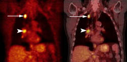 Figure 4. Scan of a lung cancer patient with a nodule in the right upper lobe. The PET scan is on the left. The picture on the right is a FUSION image of the PET and CT scan. Notice the bright yellow spot in the right upper lobe (arrow) and the 2nd spot in the hilum (arrow head). This information tells the surgeon that the disease has likely spread to this region.
By identifying unexpected distant metastases in 10-20% of patients (5), PET helps to avoid a nontherapeutic operation. PET is also attractive since the whole body can be imaged in one session. FDG-PET has been reported to be 100% sensitive for adrenal metastases (5) although PET is not accurate in identifying brain metastases due to normal uptake of FDG by the brain.
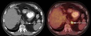 Figure 5. CT with Fusion PET image on same patient. Notice that the adrenal gland (arrow) appears as a bright yellow spot on the FUSION image. This implies the disease has spread outside the chest. In addition, metabolism may be altered by infection leading to false results in areas where histoplasmosis or granulomatous disease are common. Current recommendations require confirmation of all PET-positive mediastinal nodes with mediastinoscopy. PET is also useful in guiding mediastinal biopsy, especially when disease is identified in a nodal region not accessible by mediastinoscopy alone. Whether negative studies should be followed by mediastinoscopy is controversial and depends on surgeon preference and the institutional experience.
PET scanning provides functional information and has been found to be useful for determining the diagnosis, stage, and prognosis of lung cancer. Several studies have shown that PET is more accurate than CT for staging mediastinal lymph nodes (7). This powerful new tool is revolutionizing the way surgeons care for lung cancer patients. More accurate staging also helps the Oncologists and Radiation Therapists to plan the most appropriate treatment for patients who have inoperable disease.