Adult Breast Morphology
Location/Anatomic Boundaries
The adult breast lies between the second and sixth ribs in the vertical plane and between the sternal edge medially and midaxillary line laterally.
The average breast measures 10-12 cm in diameter, and thickness centrally is 5-7 cm. It is concentric with a lateral projection into the axilla, referred to as the axillary tail of Spence.
Structures
The adult breast consists of three major structures: skin, subcutaneous fatty tissue and breast tissue (parenchyma and stroma). The skin contains hair follicles, sebaceous glands and eccrine sweat glands.
The glandular breast is divided into 15-20 segments (lobes) that converge at the nipple in a radial arrangement. These lobes are made up of 20-40 lobules. Each lobule in turn consists of 10-100 alveoli (tubolosaccular secretory units). Collecting milk ducts, measuring approximately 2 mm in diameter, drain each segment. Between five to ten major collecting milk ducts open at the nipple into subareolar lactiferous sinuses, which are about 5-8 mm in diameter. Between the lobes of glandular tissue is subcutaneous connective tissue.
Superficial pectoral fascia envelops the breast and is continuous with the superficial abdominal fascia of Camper. The undersurface of the breast lies on the deep pectoral fascia. Cooper suspensory ligaments provide support for the breast and are fibrous bands connecting the two fascial layers. The retromammary bursa refers to a distinct space on the posterior aspect of the breast between the deep layers of the superficial fascia of the breast and the deep investing fascia of the pectoralis major.
Nipple / Areola
The epidermis of the nipple (mammary papilla) and areola is pigmented and wrinkled and consists of keratinized, stratified squamous epithelium. It is 15-60 mm in diameter. There are bundles of smooth-muscle fibers that are circumferentially arranged in dense connective tissue and are responsible for the contractile function and erection of the nipple. Two receptor-type nerve endings (Ruffini-like bodies and end bulb of Krause) are present on the nipple and are associated with the tactile reception of stretch and pressure.
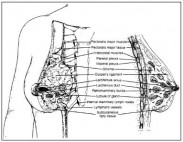
Neuronal plexuses around hair follicles in the skin peripheral to the areola are also present. The areola has no hair follicles. It has sebaceous glands (at its margin), apocrine sweat glands, and accessory areolar glands (Montgomery glands). Montgomery glands are intermediate between true mammary glands and sweat glands and open on the surface of the areola as small elevations called Morgagni tubercles. Figure 1.1 is a tangential view of the breast on the chest wall and a sectional view of the breast and chest wall.
Blood Supply of the Breast
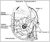
The blood supply of the breast is mostly from superficial vessels. The principal blood supply is derived from the internal thoracic (mammary) and lateral thoracic artery. Enlarged lateral branches of the anterior perforating arteries originating from the internal thoracic artery run to the breast as medial mammary arteries. The lateral mammary arteries are often multiple in origin and are derived from the lateral thoracic artery. The posterior intercostal arteries of the second and fourth intercostal spaces also give off mammary branches. The superficial veins follow the arteries and drain through perforating branches of the internal thoracic vein, tributaries of the axillary vein and perforating branches of posterior intercostal veins. The veins anastomose circumferentially around the nipple, the circulus venosus. Figure 1.2 illustrates the blood supply to the breast.
Innervation of the Breast
Sensory innervation is supplied primarily by the lateral and anterior cutaneous branches of the second through sixth intercostal nerves. Branches of the supraclavicular nerve supply a limited portion of the skin in the upper breast.
Secretory activity is under pituitary and ovarian hormonal control. Oxytocin released in the neurohypophysis stimulates myoepithelial cells that in turn stimulate contraction and release of milk from the mammary glands.
Mary L. Gemignani
Memorial Sloan-Kettering Cancer Center