Muscular Abnormalities
Congenital absence of the sternocostal head of the pectoralis major muscle and its associated abnormalities has been described earlier in this chapter. In 5% of cadavers, a sternalis muscle can be found lying longitudinally between the sternal insertion of the sternocleidomastoid muscle and the rectus abdominis muscle.
The pectoralis minor muscle is inserted into the head of the humerus and the coracoid process of the scapula in 15% of cases. Part of the tendon then passes between the two parts of the coracoacromial ligament to insert into the coracohumeral ligament.
Rarely, the axillopectoral muscle arises as a separate part of the latissimus dorsi muscle and crosses the base of the axilla superficially, then passes deep to the pectoralis major muscle to join its insertion or to continue to the coracoid process (the Langer axillary arch). This anatomic arrangement can cause compression of the axillary vessels and difficulty in orientation during axillary dissection.
Microanatomy of Breast Development
The developing breast at puberty has been described in detail by Russo and Russo as growing and dividing ducts that form club-shaped terminal end buds. Growing terminal end buds form new branches, twigs, and small ductules termed alveolar buds. Alveolar buds subsequently differentiate into the terminal structure of the resting breast, named the acines by German pathologists or the ductule by Dawson. The term alveolus is best applied to the resting secretory unit and acines to the fully developed secretory unit of pregnancy and lactation.
![]() TABLE 3. Characteristics of Human Breast Lobules. Lobules develop during the first few years after menarche. The alveolar buds cluster around a terminal duct and form type I (virginal) lobules, comprising approximately 11 alveolar buds lined by two layers of epithelium. Full differentiation of the mammary gland proceeds through puberty, takes many years, and may not be fully completed if interrupted by pregnancies.
TABLE 3. Characteristics of Human Breast Lobules. Lobules develop during the first few years after menarche. The alveolar buds cluster around a terminal duct and form type I (virginal) lobules, comprising approximately 11 alveolar buds lined by two layers of epithelium. Full differentiation of the mammary gland proceeds through puberty, takes many years, and may not be fully completed if interrupted by pregnancies.
![]() Detailed microanatomic studies of the breast have shown the presence of three distinct types of lobules. Type I lobules, previously described, are the first generation of lobules that develop just after the menarche. The transition to type II and type III gradually results from continued sprouting of new alveolar buds. The characteristics of the four lobular types are described in Table 1-3 and Table 1-4.
Detailed microanatomic studies of the breast have shown the presence of three distinct types of lobules. Type I lobules, previously described, are the first generation of lobules that develop just after the menarche. The transition to type II and type III gradually results from continued sprouting of new alveolar buds. The characteristics of the four lobular types are described in Table 1-3 and Table 1-4.
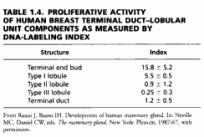 TABLE 4. Proliferative Activity of Human Breast Terminal Duct–Lobular Unit Components as Measured by DNA-Labeling Index
Microscopic Anatomy of the Adult Breast
In the immature breast, the ducts and alveoli are lined by a two-layer epithelium that consists of a basal cuboidal layer and a flattened surface layer. In the presence of estrogens at puberty and subsequently, this epithelium proliferates, becoming multilayered. Three alveolar cell types have been observed: superficial (luminal) A cells, basal B cells (chief cells), and myoepithelial cells.
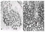 FIG. 4. A: One normal, inactive lobule from the breast of a 35-year-old woman. Alveolar cells are small and the lumina are inconspicuous. Much of the lobular space is occupied by capillaries and stroma. (Hematoxylin-eosin stain; x40.)
B: Portions of three adjacent, lactating lobules from the breast of a 25-year-old woman who was 3 weeks postpartum. There is cytoplasmic vacuolization of the alveolar cells, which have proliferated, leading to marked enlargement of the lobule. (Hematoxylin-eosin stain; x40.)
Superficial, or luminal, A cells are dark, basophilic-staining cells that are rich in ribosomes. Superficial cells undergo intercellular dehiscence, with swelling of the mitochondria, and become grouped, forming buds within the lumen. Basal B cells, or chief cells, are the major cell type in mammary epithelium. They are clear, with an ovoid nucleus without nucleoli. Where the basal cells are in contact with the lumen, microvilli occur on the cell membrane. Intracytoplasmic filaments are similar to those in myoepithelial cells, suggesting their differentiation toward that cell type. Myoepithelial cells are located around alveoli and small excretory milk ducts between the inner aspect of the basement membrane and the tunica propria. Myoepithelial cells are arranged in a branching, starlike fashion. The sarcoplasm contains filaments that are 50 to 80 nm in diameter; these myofilaments are inserted by hemidesmosomes into the basal membrane. These cells are not innervated but are stimulated by the steroid hormones prolactin and oxytocin.
M. P. Osborne: Department of Surgery, Joan and Sanford I. Weill Medical College, Cornell University
New York Presbyterian Hospital, New York, New York