Breast Changes during Pregnancy
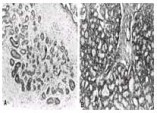
During pregnancy, marked ductular, lobular, and alveolar growth occurs as a result of the influence of luteal and placental sex steroids, placental lactogen, prolactin, and chorionic gonadotropin (Fig. 1-4B). In experimental studies, these effects are observed when estrogen and progesterone cause a release of prolactin by reducing the hypothalamic release of prolactin-inhibiting factor (PIF). Prolactin in humans is also released progressively during pregnancy and probably stimulates epithelial growth and secretion. Prolactin increases slowly during the first half of pregnancy; during the second and third trimesters, blood levels of prolactin are three to five times higher than normal, and mammary epithelium initiates protein synthesis.
In the first 3 to 4 weeks of pregnancy, marked ductular sprouting occurs with some branching, and lobular formation occurs under estrogenic influence. At 5 to 8 weeks, breast enlargement is significant, with dilatation of the superficial veins, heaviness, and increasing pigmentation of the nipple–areolar complex. In the second trimester, lobular formation exceeds ductular sprouting under progestogenic influence. The alveoli contain colostrum but no fat, which is secreted under the influence of prolactin. From the second half of pregnancy onward, increasing breast size results not from mammary epithelial proliferation but from increasing dilatation of the alveoli with colostrum, as well as from hypertrophy of myoepithelial cells, connective tissue, and fat. If these processes are interrupted by early delivery, lactation may be adequate from 16 weeks of pregnancy onward.
At the beginning of the second trimester, the mammary alveoli, but not the milk ducts, lose the superficial layer of A cells. Before this, as in the nonpregnant woman, the two-layer structure is maintained. In the second and third trimesters, this monolayer differentiates into a colostrum–cell layer and accumulates eosinophilic cells, plasma cells, and leukocytes around the alveoli. As pregnancy continues, colostrum, composed of desquamated epithelial cells, accumulates. Aggregations of lymphocytes, round cells, and desquamated phagocytic alveolar cells (foam cells) may be found in colostrum; these are termed the Donne corpuscles.
Lactation
After parturition, an immediate withdrawal of placental lactogen and sex steroid hormones occurs. During pregnancy, these hormones antagonize the effect of prolactin on mammary epithelium. Concomitant to the abrupt removal of the placental hormones, luteal production of the sex steroid hormones also ceases. A nadir is reached on the fourth to fifth day postpartum; at this time, the secretion of PIF from the hypothalamus into the hypothalamoadenohypophyseal portal system decreases. This reduction in PIF secretion allows the transmembrane secretion of prolactin by pituitary lactotrophs. Sex steroid hormones are not necessary for successful lactation, and physiologic increases, such as may occur with postpartum ovulatory cycles, do not inhibit it.
Prolactin, in the presence of growth hormone, insulin, and cortisol, converts the mammary epithelial cells from a presecretory to a secretory state. During the first 4 or 5 days after birth, the breasts enlarge as a result of the accumulation of secretions in the alveoli and ducts. The initial secretion is of colostrum, a thin, serous fluid that is, at first, sticky and yellow. Colostrum contains lactoglobulin, which is identical to blood immunoglobulins. The importance of these immunoglobulins is unknown; many maternal antibodies cross the placenta, transferring passive immunity to the fetus in utero. Fatty acids, such as decadienoic acid, phospholipids, fat-soluble vitamins, and lactalbumin, in colostrum have considerable nutritional value. After colostrum secretion, transitional milk and then mature milk are elaborated.
Mechanisms of Milk Synthesis and Secretion
The effects of prolactin are mediated through membrane receptors in the mammary epithelial cells. The release of prolactin is maintained and stimulated by suckling, as is the release of corticotropin (adrenocorticotropic hormone). The mammary cells are cuboidal, depending on the degree of intracellular accumulation of secretions. The DNA and RNA of the nuclei increase, and abundant mitochondria, ribosomes, and rough endoplasmic reticulum, with a prominent Golgi apparatus, are apparent in the epithelial cells. Complex protein, mild fat, and lactose synthetic pathways are activated, as are water-ion transport mechanisms. These processes are initiated by the activation of hormone-specific membrane receptors. Changes in cyclic adenosine monophosphate stimulate milk synthesis through the induction of messenger and transfer RNA. Prolactin stimulates cyclic adenosine monophosphate–induced protein kinase activity, resulting in the phosphorylation of milk proteins. Polymerase activity and cellular transcription are enhanced.
Large fat vacuoles develop and move toward the apex of the cell. At the same time, the nucleus also moves toward the apex. As the water intake of the cell increases, longitudinal cellular striations may be observed. Ultimately, the vacuoles pass from the cell along with part of the cell membrane and cytoplasm; the apical cell membrane reconstitutes as secretion takes place.
Enhanced activity occurs during suckling. Fat is secreted chiefly through an apocrine mechanism, lactose is secreted through a merocrine mechanism, and the secretion of proteins occurs as a result of a combination of mechanisms. Ions enter the milk by diffusion and active transport. Relatively little holocrine secretion is thought to take place. The end result of secretion and subsequent intraductal dilution of extracellular fluid is milk, comprising a suspension of proteins - casein, b-lactalbumin, and b-lactoglobulin - and fat in a lactose-mineral solution. The white appearance of milk is due to emulsified lipids and calcium caseinate, whereas the yellow color of butterfat is due to the presence of carotenoids.
Mechanisms of Milk Ejection
The removal of milk by suckling is aided by active ejection. Sensory nerve endings in the nipple-areolar complex are activated by tactile stimuli. Impulses pass by way of sensory nerves through the dorsal roots to the spinal cord. In the spinal cord, impulses are relayed through the dorsal, lateral, and ventral spinothalamic tracts to the mesencephalon and lateral hypothalamus. Inhibition of PIF secretion permits the unimpeded secretion of prolactin from the anterior pituitary. Simultaneously, through a different pathway in the paraventricular nucleus, the synthesis of oxytocin occurs. Oxytocin is released from the posterior pituitary neurovesicles by impulses traveling along the neurosecretory fibers of the hypothalamoneurohypophyseal tract. Oxytocin released into the systemic circulation acts on the myoepithelial cells, which contract and eject milk from the alveoli into the lactiferous ducts and sinuses. This phenomenon is specific to oxytocin, and changes in intramammary ductal pressures of 20 to 25 mm Hg may be observed in relation to peak blood levels. Oxytocin also acts on the uterus and cervix to promote involution. This effect may be stimulated by cervical dilatation and by vaginal stretching through the ascending afferent neural pathways (Ferguson reflex).
Complex neuroendocrine interactions determine normal lactation. An appreciation of these mechanisms is essential to the understanding of abnormalities and to the treatment of problems of lactation.
Menopause
Declining ovarian function in late premenopause through the menopause leads to regression of epithelial structures and stroma. The duct system remains, but the lobules shrink and collapse. The last structures to appear with sexual maturity are the first ones to regress.
M. P. Osborne: Department of Surgery, Joan and Sanford I. Weill Medical College, Cornell University
New York Presbyterian Hospital, New York, New York