Axilla - Anatomy of the Breast and Axilla
1. The supreme thoracic artery supplies the thoracic wall over the first and second intercostal spaces.
2. The thoracoacromial trunk divides into the acromial, clavicular, deltoid and pectoral branches.
3. The lateral thoracic artery passes along the lateral border of the pectoralis minor on the superficial surface of the serratus anterior muscle. It also supplies the lateral mammary branches. The pectoral branches of the thoracoacromial trunk as well as the lateral thoracic artery supply the pectoralis major and minor.
4. Both the anterior and posterior circumflex humeral arteries supply the upper arm and contribute to the collateral circulation around the shoulder.
5. The subscapular artery is the largest branch within the axilla. It is closely associated with the central and subscapular lymph node groups. It branches into the subscapular circumflex and the thoracodorsal artery. The thoracodorsal crosses and supplies the subscapularis. It also gives branches to the serratus anterior and the latissimus dorsi. Figure 1.4 shows the axillary artery and the brachial plexus in situ.
Axillary Vein
The axillary vein begins at the union of the basilic and brachial veins and terminates at the first rib as the subclavian vein. It lies medially and partly overlaps the axillary artery. It receives tributaries (usually paired veins) that correspond to the branches of the axillary artery and the cephalic vein. The cephalic vein lies in the groove between the deltoid and pectoral muscles and pierces the clavipectoral fascia to join the axillary vein.
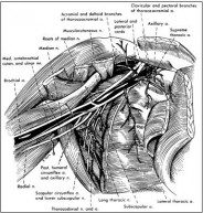 Fig. 1.4. Axillary artery and brachial plexus. The axillary artery, veins, its branches and the brachial plexus are shown in situ. The lymph nodes, veins and the pectoral nerves have been removed. Reprinted with permission from: Rosse C, Gaddum-Rosse.
P. Hollinshead’s Textbook of Anatomy. 5th ed. 1997; 215. ©1997 Lippincott-Raven
Brachial Plexus
The brachial plexus is formed by the union of the ventral rami of C5-C8 and of most of the ventral ramus of the first thoracic nerve. Often a small part of the fourth cervical nerve also joins the plexus. These nerve eventually form trunks, anterior and posterior divisions and cords. In the axilla, the anterior and posterior divisions form three cords. The three cords of the brachial plexus are named according to their position in relation to the axillary artery. Table 1.4 lists the cords of the brachial plexus, their divisions and terminal branches.
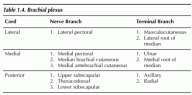
- The lateral pectoral nerve supplies the pectoralis major and gives a branch that communicates with the medial pectoral nerve. It pierces the clavipectoral fascia with the thoracoacromial artery and enters the deep surface of the clavicular head of the pectoralis major.
- The medial pectoral nerve passes between the axillary artery and vein to enter the deep surface of the pectoralis minor and continues into the pectoralis major. It is the main nerve innervation to the pectoralis minor.
- The thoracodorsal runs downward, crossing the lateral border of the scapula and the teres major to enter the costal surface of the latissimus dorsi.
- The long thoracic nerve is located on the medial wall of the axilla on the serratus anterior. It arises from the C5-C7 roots and enters the axilla through the cervicoaxillary canal. Injury to this nerve results in paralysis to part or all of the serratus anterior. The functional deficit is inability to raise the arm above the level of the shoulder.
- The intercostobrachial nerve is formed by the joining of a lateral cutaneous branch of the second intercostal nerve with the medial cutaneous nerve of the arm. This nerve supplies the skin of the floor of the axilla and of the upper medial aspect of the arm. A second intercostobrachial nerve may also form an anterior branch with the third lateral cutaneous nerve of the arm. Injury will cause numbness of the floor of the axilla and of the medial aspect of the arm.