Health Centers > Cancer Health Center > Esophageal Cancer Treatment
Esophageal Cancer Treatment
The next step following the diagnosis of esophageal cancer is to determine whether the patient is a candidate for major curative surgery. Unfortunately, over 60% of patients with esophageal cancer are not candidates for surgery at the time of presentation due to the advanced stage of disease or significant comorbidity that would result in high perioperative mortality. The combination of a thin wall, absence of a serosa, and an extensive lymphatic drainage system facilitates early regional and distant metastases in esophageal cancer. Extensive medical problems, most commonly severe cardiopulmonary disease, are contraindications to surgery. These factors demand that treatment of esophageal cancer be based on a systematic evaluation (Table 18-5). Cancer staging is generally not required for patients who are not candidates for surgery.
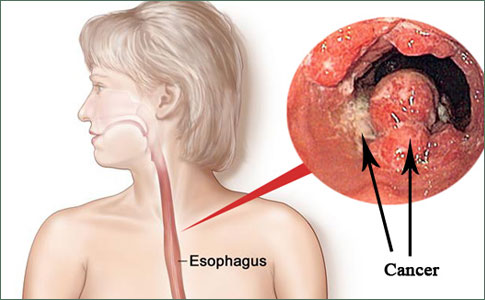
Esophageal cancer
Demographics & Epidemiology
Etiology
Natural History
¬ Clinical Presentation
¬ Complications
¬ Prognostic Factors
Diagnosis
Staging Techniques
Differential Diagnosis
Treatment
Surgery
¬ General Considerations
¬ Surgical Approach
Palliation Therapy
¬ Radiation Therapy
¬ Chemotherapy
¬ ¬ Single agent
¬ ¬ Combination chemotherapy
¬ ¬ Combined-modality therapy
¬ Endoscopic Therapy
¬ ¬ Dilation
¬ ¬ Ablation
¬ ¬ Photodynamic therapy (PDT)
¬ ¬ Endoscopic mucosal resection (EMR)
¬ ¬ Esophageal prostheses
Prognosis
Surgery
Surgical therapy has long been the preferred approach for both cure and palliation of patients with resectable tumors. Patients in the subgroup with limited local spread (T1-T2) and no regional nodal involvement are potentially curable by surgery. Patients who are deemed surgical candidates based on their good medical condition should undergo thorough staging to determine if they have curable disease, incurable but resectable disease, or unresectable disease. Resectable tumors are characterized by the absence of extension into mediastinal structures and the absence of nodal or organ metastases. Direct invasion of the aorta, bronchi, pleura, or laryngeal nerve or distant organ metastases are evidence of nonresectable disease. Preoperative radiation with or without chemotherapy may sometimes downstage the cancer to a resectable or potentially curable stage.
Prior to surgery, it is important to confirm that the patient has sufficient cardiopulmonary reserve. Respiratory function is best assessed by the forced expiratory volume (FEV1), which, ideally, should be 2 L or more. Any patient with an FEV1 of less than 1.25 L is a poor surgical candidate due to the high risk of respiratory insufficiency postoperatively. Cardiac reserve should be assessed and a resting ejection fraction of less than 40% is an ominous finding.
Perioperatively, nutritional support should be provided to improve postoperative complications and recovery. A poor nutritional status affects the host resistance to infections and impairs anastomotic and wound healing. As oral intake is usually inadequate in patients with advanced disease, a feeding jejunostomy tube is the most reliable and safest method for nutritional support in those with a functional small bowel. A gastrostomy is inadvisable for these patients because it may interfere with the use of the stomach for reconstruction. The jejunostomy also minimizes the danger of regurgitation into the pharynx and possible aspiration. Total parenteral nutrition may also be indicated for some patients.
Anal Cancer: Strategies in Management
The management of anal cancer underwent an interesting transformation over the last two decades.
The surgical options available for esophageal tumor resection include (1) transhiatal, (2) combined right thoracic and abdominal (Ivor Lewis), (3) left thoracoabdominal, and (4) en bloc, either two field or three field. In a simple esophagectomy, whether by the transhiatal or the transthoracic route, there is no specific attempt to remove lymph node tissues in the mediastinum or upper abdomen. Cure is thus uncommon and occurs only by chance. En bloc resection involves a radical esophagectomy to include mediastinal, upper abdominal (two field), and/or cervical (three field) lymphadenectomy.
Transhiatal esophagectomy is currently the preferred surgical approach for palliation of esophageal cancer independent of its location. In patients with distal esophageal cancer, transhiatal esophagectomy may be curative when adequate inspection of the paraesophageal cancer tissue is provided by the abdominal incision. The operation consists of abdominal and cervical incisions and a cervical gastroesophageal anastomosis. This procedure does not allow visual inspection of the mediastinal bed, which theoretically is necessary to ensure removal of the locally invasive tumor, although it offers a slightly better 3-year survival and operative mortality than the transthoracic approach (25% versus 20% and 5% versus 10%, respectively).
En bloc resection remains the definitive surgical cure for esophageal cancer. The operation consists of removal of a tissue block completely surrounded by normal tissue. Two or three fields of lymphatic resection are included depending upon the tumor location: upper abdominal celiac and splenic nodes (field one), infracarinal posterior mediastinal nodes (field two), and upper mediastinal and cervical (field three). Recent studies encouraged inclusion of the surrounding mediastinal pleura, the azygos vein, the thoracic duct, and possibly the pericardium at the time of surgery to improve the overall survival. The overall 5-year survival of en bloc resection approaches 40% although the operative mortality still ranges from 5 to 10%.
Benign Esophageal Tumors
Benign esophageal tumors
General Considerations
Clinical Findings
¬ Symptoms and Signs
¬ Imaging
Treatment
References
Locally advanced tumors (T1N1, T2N1, T3N0, and T3N1) are resectable but incurable. These tumors are associated with a high recurrence rate following surgery. The optimal intervention in these patients remains controversial, and practices vary widely depending on the local surgical expertise and the assessment of the patient's preexisting medical condition.
Palliation Therapy
As the majority of patients present with incurable disease, pallative therapy remains the mainstay of treatment options for esophageal cancer. The options currently available include radiation therapy, chemotherapy, endoscopic dilation and ablation (chemical injection, electrocautery, argon plasma coagulation, and laser), photodynamic therapy, and esophageal prostheses/stents. They are all palliative procedures employed as adjuvant therapy to surgery or for patients who are not considered surgical candidates. The treatment again varies according to availability and local expertise, patient preference, and cost.
External beam radiation therapy alone provides reasonable palliation for esophageal cancer, especially for those who are medically unable to undergo surgery or chemotherapy. At the end of treatment, radiotherapy achieves palliation of dysphagia in 70-90% of patients. The 5-year survival curves are similar to those for surgery for patients with a comparable cancer stage (5-10%). It is recommended that for patients treated with curative intent, radiation therapy should be limited to tumors 10 cm with no evidence of distant metastasis. Contraindications to radiotherapy include tracheal or bronchial involvement, cervical esophagus location of the tumor, or stenosis that cannot be bypassed. The major complications of radiation therapy in esophageal cancer are airway fistulas (10-15%) and esophageal strictures (20-40%).
In an attempt to improve its effectiveness, radiotherapy has been given in a hyperfractionated manner. Several recent studies employing continuous hyperfractionated accelerated radiotherapy demonstrate a slightly improved median survival and prolonged relief of dysphagia, although there is no significant improvement in the 5-year survival rates. In addition, many studies have looked into the role of radiation therapy as an adjuvant to surgery to improve local tumor control and survival. Several randomized control studies comparing the benefit of preoperative and/or postoperative radiation in addition to surgery demonstrated disappointing results. There is no additional survival benefit, and there seems to be a higher rate of operative complications following radiation.
By combining external beam radiation with intraluminal irradiation using cobalt-60, cesium-137, or iridium-192, it is possible to increase the dose of radiation to the tumor without significantly increasing the dose of radiation to normal tissue. A major limitation of intraluminal irradiation is the effective treatment distance. Initial studies have demonstrated promising results with improved median survival and 5-year survival, and more clinical trials are underway.
1. Single agent - Despite the increasing choice of agents, chemotherapy alone has been of little benefit to patients with esophageal cancer. The most commonly employed classes of agents for treating esophageal cancer include (1) antibiotics - bleomycin and mitomycin C, (2) antimetabolites - 5-fluorouracil (5-FU) and methotrexate, (3) alkaloids - vindesine and vinorelbine, (4) platinum analogs - cisplatin, carboplatin, and oxaliplatin, (5) taxanes - paclitaxel and docetaxel, and (6) topoisomerase inhibitors - etoposide and irinotecan. The clinical response rates for most single agents are very poor (5-15%), although cisplatin and the taxanes are exceptions and have been the focus of most combination chemotherapy. The response durations are also very brief, ranging from 2 to 4 months.
2. Combination chemotherapy - Combination chemotherapy has typically demonstrated better clinical response rates than single agent chemotherapy. Most trials of combination chemotherapy are based on cisplatin, as it alone has a response rate of 20-25%. In general, the cisplatin-based combination chemotherapy has yielded a response rate of 25-35%. The results are even more impressive in locoregional disease, yielding 45-75%. Unfortunately, the higher response rates do not translate into improved response duration or improved survival. In addition, the higher response rate of combination chemotherapy needs to be balanced against a higher systemic toxicity. To date, there is no role for chemotherapy, single agent or combination, as an adjuvant to surgery.
3. Combined-modality therapy - As primary management, combined-modality therapy of chemoradiation has achieved better median survival and 5-year survival when compared with radiation alone. In addition, there is much current interest in the role of chemoradiation as induction therapy prior to surgery. Theoretically, induction chemoradiation therapy has several advantages over primary surgery alone or postoperative chemoradiation. As the local blood circulation is not yet disrupted by surgical dissection, preoperative chemotherapy should result in better drug delivery to the tumor. Preoperative treatment also allows for identification of patients who may in turn benefit from further postoperative therapy if needed. In addition, concurrent chemoradiation can take advantage of the radiation-sensitizing properties of many chemotherapeutic agents (eg, paclitaxel, 5-FU, and cisplatin), resulting in a synergistic antitumor effect. Distant control should be enhanced as remote micrometastases are treated early in the course instead of having to wait for postsurgical recovery. Preliminary results from recent studies are encouraging, and many trials are currently underway to define the role of chemoradiation further.
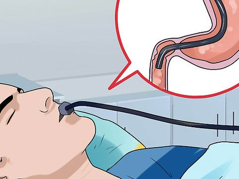
Whereas endoscopy has mainly been used to diagnose cancers, new technologies such as photodynamic therapy and endoscopic mucosal resection have provided endoscopic treatments with the potential of curing early stage tumors of the esophagus.
1. Dilation - Esophageal dilation is most commonly performed with either expandable through-the-scope balloons or wire-guided polyvinyl bougies under fluoroscopy. A small percentage of patients can be successfully dilated to allow for soft diet consumption. However, the benefit from dilation is usually of short duration, and other methods are usually required for more prolonged symptom relief. One of the major complications of esophageal dilation is perforation.
2. Ablation - Tumor ablation treatments available for palliation include photodynamic therapy, chemical sclerosant injection, monopolar and bipolar electrocautery, argon plasma coagulation (APC), and neodymium:yttrium-aluminum-garnet (Nd:YAG) laser. The simplest and least expensive intervention for esophageal cancer ablation is the injection of a chemical sclerosant during endoscopy. Absolute alcohol is the most widely employed chemical agent. The method has been shown to be capable of results similar to laser therapy. The major problem with chemical injection relates to a lack of control as the sclerosant tracks along the tissue planes, causing damage to normal tissue and, sometimes, perforations. Patients may experience temporary worsening of symptoms until adequate tumor necrosis occurs.
Monopolar and bipolar electrocautery are falling out of favor and are rarely used as it is difficult to control the depth of treatment. A newer method, argon plasma coagulation, uses ionized argon gas to convey electrical energy to achieve thermal desiccation of tumor tissue. Unfortunately, the effect is generally superficial, and it is less efficient in relieving dysphagia in advanced esophageal cancer than the tissue ablation achieved using lasers.
High-power Nd:YAG laser can provide palliation of dysphagia by coagulating and vaporizing malignant tissue under endoscopic control. Tumors amenable to laser therapy are exophytic or polypoid, preferably located in a straight segment of the esophagus such as in the mid-esophagus or lower esophagus, and shorter than 5 cm. Multiple endoscopic laser treatment sessions may be required to reduce the size of the intraluminal tumor to improve swallowing. Periodic follow-up is performed to reduce any recurrent intraluminal tumor growth. Although more expensive, laser treatment is more widely available.
3. Photodynamic therapy (PDT) - PDT has emerged as an attractive palliative treatment for esophageal cancer and its complications. PDT has been successful in reducing tumor bulk and in opening the esophageal lumen in patients with complete obstruction, a situation in which Nd:YAG therapy was considered too risky. PDT has also been utilized as a salvage therapy in patients whose stents have failed because of tumor ingrowth/overgrowth.
The treatment begins with an intravenous injection of a photosensitive chemical, porfimer sodium (Photofrin). It is administered at a dose of 2 mg/kg of body weight and preferentially concentrates in the tumor tissue. After 40-50 hours following the injection, the area of the esophageal cancer is exposed to a red light at a wavelength of 630 nm, delivered from a continuous-wave dye laser via an optical fiber diffuser for a total cumulative light dose of 300 J/cm. The red light has been chosen for greatest depth of penetration (5 mm). The process initiates a photochemical reaction and the effect takes place over the ensuing hours to days, ultimately resulting in necrosis of the tumor. Improvement in the dysphagia is usually noted within 5-7 days, although some patients may experience worsening of dysphagia initially due to local tissue inflammation and edema. The major issue with PDT is retention of the photosensitive dye in the skin (up to 6 weeks), which requires patients to avoid direct sun exposure or risk severe sunburn. Other complications following PDT include fever, leukocytosis, nausea, and pleural effusion. Severe complications, which fortunately are uncommon, include atrial arrhythmias, stricture formation, hemorrhage, and perforation. The advantage of PDT in early esophageal cancer (T1 or T2 disease) is the overall greater than 80% cure rate.
4. Endoscopic mucosal resection (EMR) - The development of EMR, or mucosectomy, was sparked by the need to treat superficial flat and polypoid neoplasms of the mucosa of the gastrointestinal (GI) tract with minimally invasive procedures. Long-term studies have demonstrated that EMR outcomes are similar to those of surgery, which has led to acceptance of EMR as a standard treatment, especially in early stage GI cancers. The availability of endoscopic ultrasound to determine the depth of tumor invasion and lymph node metastases as well as chromoendoscopic techniques to reveal tumor borders otherwise not visible without staining further facilitate the ease and use of EMR.
Numerous EMR techniques have been described, such as injection and snare cautery, injection with precut, EMR with cap, or EMR with band ligation. Nevertheless, the general principles are the same. EMR involves expansion of the submucosal layer and lifting of the mucosa to allow for a safe longitudinal resection. Injection of an expansion solution (typically normal saline) into the submucosa creates a bleb and increases the distance between the mucosa and muscularis propia. This lifting of the mucosa is essential to prevent transmural burning or perforation. A snare is placed over the base of the "neopolyp" and the mucosa is resected with electrocautery. The cancerous lesion is removed en bloc and allows for a complete detailed histopathologic analysis.
In esophageal cancers, EMR has been applied mainly for squamous cell carcinoma and much less for Barrett's esophagus-related adenocarcinoma. EMR is indicated when the lesion is superficial and without evidence of lymph node metastasis. Although conventional EUS is accurate in determining tumor depth and lymph node metastasis of large or bulky lesions, it is less precise for small, flat, or depressed tumors. Therefore, in these instances, high-frequency ultrasound probes have been recommended. The application of Lugol's solution further assists in visual distinction between a cancerous lesion and normal mucosa (squamous cell carcinoma or dysplasia does not stain). Once the lesion is identified and staged, approximately 20 mL of saline is injected into the submucosa, causing more than half-circumferential mucosal lifting. EMR can then be safely performed. EMR should be avoided if the lesion cannot be lifted by submucosal saline injection (nonlifting sign), which does occur when a tumor has indeed invaded the muscularis propia or when there has been fibrosis due to prior polypectomy. There is no consensus on the maximal lesion size suitable for EMR in esophageal cancer. However, it is recommended that the lesion should be less than 3 cm in height and should not exceed one-third of the esophageal circumference in width to avoid the late complication of stenosis. For larger lesions, other alternatives, such as photodynamic therapy, should be considered.
The major complications of esophageal EMR are bleeding, perforation, and stenosis. Bleeding during EMR is almost always controllable by injection of a low-concentration epinephrine-saline solution, thermal coagulation, or endoscopic clipping. Large perforations invariably require immediate surgery, although small perforations may be manageable with more conservative measures. Stenosis can occur when circumferential resection has been attempted.
5. Esophageal prostheses - Placement of esophageal prostheses ("stents") is another appealing method for the palliation of malignant strictures and provides effective relief of dysphagia in most cases. The prostheses are usually inserted surgically or endoscopically with fluoroscopic guidance. A wide variety of stents have been developed and modified over the years that provide good mechanical support for maintaining esophageal lumen patency and reduce complications such as stent migration and tumor ingrowth. At present, the expandable metal stents are the most widely applied form of prostheses. The principal merits of such stents are the ease of insertion and the remarkably low risk of esophageal perforation. Covering the stent with a polymer sheet effectively reduces tumor ingrowth and provides for treatment of tracheoesophageal fistulas, while the outer flange diameters have been increased, thereby making a funnel-shaped stent that adheres to the esophageal wall to significantly lower the rate of migration. Early complications of expandable metal stents have included incomplete expansion, perforation, bleeding, and pain. Late complications include tumor overgrowth/ingrowth, ulceration, food impaction, and stent migration. Patients who have received prior radiation and chemotherapy are more prone to develop complications. Stents that cross into the gastric cardia may cause significant gastroesophageal reflux that can usually be controlled with proton-pump inhibitors; however, a new stent has been developed that prevents reflux via a valve mechanism. In cases in which patients developed an airway fistula, such as bronchoesophageal or tracheoesophageal fistula, stent placement is the optimum therapy. Furthermore, a second stent may be placed in the airway if there is significant stenosis causing dyspnea.
Prognosis
Despite the widespread use of endoscopy, significant advances in surgical techniques and neoadjuvant chemoradiation therapy, and improvements in postoperative care, the prognosis for patients with esophageal cancer remains poor. The reported overall 5-year survival rates are at best 10-15%. Delayed clinical manifestations and rapid intramural invasion and distant metastases account for the poor prognosis of this GI malignancy. Patients with an early stage of disease carry a better prognosis. For patients with T1 or T2 disease and no nodal involvement, the 5-year survival rate is greater than 40%. On the other hand, patients with T3 or T4 lesions have a 5-year survival of less than 25%.
Stage 0, I, and II tumors are considered resectable for cure. The 5-year survival for such patients who are sufficiently fit to undergo surgery ranges from greater than 85% for stage 0, to 50% for stage I, to 40% for stage II. On the other hand, stage III tumors are rarely resectable for cure, and stage IV cancers are considered incurable and nonresectable by most clinicians. The presence or absence of nodal involvement also has a significant prognostic impact. The 5-year survival for N0 disease is over 70%, whereas N1 disease is associated with a survival near 40%, independent of the T classification.