Infectious Diseases (Except Sexually Transmitted Diseases)
Vulvo-Vaginitis
The prepubertal vulva is thin, delicate, and susceptible to trauma, infection and irritation as it lacks labial fat pads and pubic hair. The anus is anatomically closer to the introitus, allowing contamination with feces and fecal pathogens.
The unestrogenized vaginal mucosa is thin and atrophic, has a neutral pH, and is an excellent medium for bacterial growth. In addition, hygiene in prepubertal children is frequently substandard as maternal supervision diminishes with age. The distinction (by history and clinical examination) between vulvitis and vaginitis has to be made. In vulvitis, dermatologic lesions involve exclusively the vulva and there is no vaginal discharge; often symptomatic treatment and advice will be enough. In cases of vaginitis, there are both clinical vulvo-vaginal manifestations and a vaginal discharge and a sexually transmitted disease has to be excluded.
Vulvitis without vaginal involvement is very common in young girls.
Symptoms include pruritus and urinary and/or anal signs. Clinical examination often shows an isolated erythema, without other dermatologic lesions. In these cases, there is no need for bacteriologic swabs as they are related to poor hygiene, irritants, and sometimes pinworms. Recommendations for improved hygiene and avoidance of irritation, in association with treatment of pinworms, are sufficient measures.
Vulvo-vaginitis with a discharge is most likely due to an infection or a retained foreign body. In this case, it is very important to clarify whether there is sexual contamination (sexually active teenagers or children who are being abused): a pertinent history and a clinical examination have to be very carefully conducted.
Any discharge must be fully investigated and samples taken for wet smears to screen for Trichomonas and Candida as well as for Gram staining and bacterial cultures. In prepubertal girls, pathogenic organisms include Streptococcus pyogenes (group A hemolytic streptococci), Haemophilus influenzae, Streptococcus pneumoniae, Staphylococcus aureus, Neisseria meningitidis, Shigella, Yersinia. Candida vulvo-vaginitis may occur in post-pubertal girls but is very rare in early infancy. Sexually transmitted disorders of bacterial origin include Trichomonas, gonorrhea and Chlamydia infections. In case of persistent vulvo-vaginitis with negative bacterial cultures, a vaginoscopic examination should be performed under anesthesia to rule out a retained foreign body.
Perineal Dermatitis
Bullous impetigo is characterized by the formation of large flaccid bullae arising from normal skin. The bullae rupture leaving red denuded round areas with honey-colored crusts. Those lesions spread rapidly and can quickly involve the thighs, buttocks, and abdomen. The causative agent is often S. aureus phage type II, which produces an epidermolytic toxin. Treatment is topical and general antibiotherapy.
Perianal dermatitis (fig. 5) induces a superficial, perianal, welldemarcated rim of erythema sometimes in association with a vulvo-vaginal erythema.
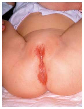
Fig. 5. Perineal streptococcal dermatitis.
Symptoms range from perianal pruritus and tenderness to abdominal pain and rectal bleeding. In girls, clinical symptoms include dysuria, pruritus, tenderness, and vaginal discharge. Clinical pharyngitis may be present.
Microbacterial cultures will grow group A b-hemolytic streptococci from perineal samples; streptococci may also be present in pharyngeal samples.
Treatment is oral penicillin V for at least 3 weeks. S. aureus has also been proven to be the cause in this disease.
Recurrent toxin-mediated perineal erythema. The hallmark of this disease is a strikingly diffuse macular erythema in the perineum occurring abruptly after a bacterial pharyngitis. Oral mucosal changes, such as strawberry tongue, as well as erythema, edema, desquamation of the hands and feet during convalescence, are usually present as well. Systemic signs are absent.
Recurrences are frequent; culture of the pharynx during acute episodes reveals toxin-producing S. aureus or S. pyogenes.
Viral Infections
Condylomata acuminata are anogenital warts caused by a human papillomavirus (HPV) infection. Most commonly, they are caused by HPV types 6, 11, 16 or 18. Sometimes, manual transmission causes condylomata with type 2 HPV. Epidemiologic and experimental inoculation studies suggest that the incubation period for HPV is 1–20 months, but latency periods of at least 2 years are suspected. The majority of condylomata acuminata in children younger than 3 years is due to vertical transmission at birth. Non-sexual transmission includes: hand-genital contact via an infected caregiver, non-sexual intimate behavior, and inadequate hygiene (contaminated towels, etc.). The possibility of sexual transmission must be considered and assessed for all children presenting with a condyloma. This includes directed medical and social history and physical examination of the child.
This also includes history of anogenital warts in the family and caregivers. Clinical examination of family members is necessary to look for warts. Condylomata acuminata present as white or flesh-colored, papilloma- like, hyperkeratotic, sharply demarcated, and non-confluent lesions. In children, they are usually perianal and localized preferentially or exclusively on the skin, not on the mucosa. As spontaneous resolution occurs quite often, non-intervention is a reasonable approach in the management of condylomata in children. If necessary, topical non-aggressive treatment should be instituted (podophillotoxin, salicylic acid, imiquimod, etc.); laser treatment or surgical excision and cauterization under general anesthesia may also be offered.
Molluscum contagiosum is caused by a poxvirus and may be localized in the genital area. Each lesion, from 1 to 10mm, is a dome-shaped papule, fleshcolored or pearly, with an umbilicated center. As it is a self-limited disease, and as treatment is often painful, non-intervention may be an alternative. Among numerous interventions, curettage or cryotherapy may be offered.
Herpes and zoster virus infection may occur in perineal regions. Genital HSV infections are rare in children and sexual contamination should be considered in this localization.
Infestations with Pinworms, Scabies and Lice
Pruritus is quite a frequent symptom in childhood and it is important to keep in mind that vulvar symptoms may be caused by anal pathologies because the anus is anatomically closer to the introitus. The diagnosis of pinworms is suggested when there is nocturnal anal pruritus; the diagnosis can be made by visualizing the worms or by applying transparent adhesive tape to the perianal skin to look for microscopic eggs. Systematic treatment is a good choice, including a single dose of oral pyrantel pamoate, 11 mg/kg, or mebendazole, 100 mg, for all household members. Both treatments may be repeated 2 weeks later. In the perineal area, scabies nodules are localized in the labia majora; eczematous lesions are concentrated on the hands, abdominal wall, and axillary folds. Pubic lice are rare in childhood.
Revision date: June 18, 2011
Last revised: by Janet A. Staessen, MD, PhD