Hemorrhoids
Hemorrhoids are masses of normal vascular tissue in the anal canal and are often the source of a variety of symptoms including bleeding, anal pruritus, prolapse, and anorectal pain due to thrombosis. The prevalence of hemorrhoids is equal in both sexes, peaking between the ages of 45 and 65 years (3). They arise from a plexus or a “cushion” of dilated veins originating from the superior and inferior hemorrhoidal veins, which are located in the submucosal layer in the lower rectum.
Hemorrhoids are classified as internal or external, based on whether they arise above or below the dentate line, and often coexist (4). Internal hemorrhoids arise from the superior (internal) hemorrhoidal vascular plexus, and their primary locations are the 3, 6, and 9 o’clock positions corresponding to the end branches of the middle and superior hemorrhoidal veins. External hemorrhoids are dilations of the inferior (external) hemorrhoidal plexus and lie below the dentate line, covered with squamous epithelium that contains numerous somatic pain receptors. External skin tags, which represent residual excess skin associated with prior thrombosis of external hemorrhoids, are often confused with external hemorrhoids but they are not hemorrhoids.
Internal and external hemorrhoids communicate with each other and drain into the internal pudendal veins, ultimately communicating with the inferior vena cava, although they have no direct communication with the portal system.
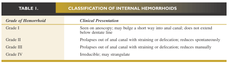
Hemorrhoids may reside in proximity to rectal varices in patients with cirrhosis but are not more common in patients with portal hypertension. There is no widely used classification system for grading external hemorrhoids, but internal hemorrhoids are graded from I to IV according to the degree to which they prolapse from the anal canal (Table I).
KEYPOINT
A thorough physical examination of the anorectum, properly done, is essential to diagnosing diseases of the rectum and anus.
Pathogenesis
The development of symptomatic hemorrhoids has been associated with increasing age, chronic diarrhea, pregnancy, pelvic tumors, prolonged sitting and straining, and possibly chronic constipation (5). Internal hemorrhoids are normal vascular cushions containing a rich arteriovenous network, which are present at birth and by partially occluding the anus contribute to continence.
These vascular cushions project into the lumen where they are subjected to downward pressure during defecation. With advancing age or aggravating conditions, the connective tissue muscle fibers that anchor the vascular cushions to the underlying sphincter mechanism become attenuated and deteriorate, allowing the hemorrhoids to slide into the anal canal and become congested, bleed, and prolapse.
Some theories postulate that symptomatic hemorrhoids arise from hypertrophy or increased tone of the internal anal sphincter and that during straining or defecation the fecal bolus forces the vascular plexus against the internal sphincter, causing hemorrhoids to enlarge and become symptomatic. These mechanisms lead to the clinical manifestations of rectal bleeding, anal pruritus, and pain associated with thrombosis.
Management of Hemorrhoids
Management approaches include conservative treatment, minimally invasive therapy, and surgical treatment (6,7). Conservative treatment is usually successful for external hemorrhoids and for most symptomatic patients where the intent is to decrease downward pressure during straining and defecation. A proper diet, use of bulk-forming agents, and avoidance of prolonged sitting are some of the conservative measures that can be taken.
Symptoms of irritation and pruritus can be usually managed with a variety of analgesic creams, hydrocortisone suppositories, and warm sitz baths.
K E Y P O I N T
Based on recent studies and meta-analysis, the optimal therapy for symptomatic grade I to III hemorrhoids refractory to conservative therapy is rubber band ligation. This technique is effective and inexpensive, requires no anesthesia, and only rarely incurs significant complications.
Analgesic creams and hydrocortisone topical therapy should not be used for longer than 7 to 10 days because side effects such as contact dermatitis or mucosal atrophy can occur. Sitz baths should be taken in warm water 2 or 3 times per day; their effectiveness may be, in part, related to relaxation of the internal anal sphincter. Thrombosis of external hemorrhoids is due to organization and resorption of clot after an acute attack and can persist despite conservative therapy. A thrombosed external hemorrhoid can cause excruciating pain, and the use of oral and topical analgesics, stool softeners, and sitz baths may provide temporary relief until resolution occurs.
Minimally invasive interventions are used for treating internal hemorrhoids and to fix the vascular cushions underlying the anal sphincter to decrease sphincter pressure. The principal approach is to remove or cause sloughing of excess hemorrhoidal tissue; the subsequent process of healing and scarring fixes the residual tissue to the anorectal muscular ring.
These therapies are often performed in an outpatient setting by an experienced anorectal surgeon and include rubber band ligation, sclerotherapy, infrared or laser photocoagulation, bipolar diathermy, cryosurgery, and dilation of the internal anal sphincter.
Based on recent studies and meta-analysis, the optimal therapy for symptomatic grade I to III hemorrhoids refractory to conservative therapy is rubber band ligation (6,7). This technique is effective and inexpensive, requires no anesthesia, and only rarely incurs significant complications.
Successful ligation leads to thrombosis of the internal hemorrhoid and the subsequent development of localized submucosal scarring. Only a single column of hemorrhoids is treated in a session to reduce the complications associated with tissue necrosis. Complications may include severe pain from misapplication of the rubber band below the dentate line or from associated anorectal spasm.
The rubber bands must be placed at least 5 mm above the dentate line to avoid somatically innervated tissue. Many patients will complain of a sense of “tightness” after the procedure; such discomfort is often alleviated by a warm sitz bath and the prevention of constipation through the use of bulk-forming fiber agents. Other complications may include:
- delayed rectal hemorrhage due to ulceration and mucosal sloughing occurring 4 to 7 days post-procedure as the rubber band falls off;
- thrombosis of hemorrhoidal tissue distal to the band ligation, causing a painful, palpable mass;
- development of severe anorectal pain, fever, or foul-smelling purulent seepage or drainage, which may signal the onset of a localized infection or abscess; and
- fulminant sepsis.
In clinical practice, however, complications of rubber band ligation are rarely reported, with only ~8% of patients reporting side effects of anorectal pain.
Sclerotherapy involves the use of specialized needles to inject sclerosants (such as morrhuate sodium, 5% phenol, and hypertonic saline), ultimately causing an intense inflammatory reaction that leads to destruction of redundant submucosal tissue and subsequent hemorrhoidal prolapse.
Sphincter dilation is performed under general anesthesia whereby the internal anal sphincter is force-fully dilated followed by daily anal dilation for several weeks. Although 80% of patients have good results, this procedure can lead to fecal incontinence due to damage of the anal sphincter and has the added disadvantages of complications related to the use of general anesthesia and high cost of the procedure.
Infrared or laser photocoagulation, bipolar diathermy, and cryosurgery are less widely employed. These therapies, which are usually reserved for grades I and II hemorrhoids, are expensive and have more side effects and increased frequency of recurrence.
Surgical management is generally reserved for patients with continued symptoms despite the application of conservative or minimally invasive procedures, or as the initial treatment of choice in cases of symptomatic grade IV hemorrhoids or in patients with strangulated internal hemorrhoids.
The most common technique is a closed hemorrhoidectomy in which an elliptical incision is made on the external hemorrhoidal tissue and extended proximally across the dentate line to the superior extent of the hemorrhoidal column (8,9). Usually 3 hemorrhoidal columns are treated at a time, and the defect is closed with a continuous absorbable suture. This procedure has a 95% success rate and a low rate of wound infection.
Open hemorrhoidectomy is recommended by some anorectal surgeons to reduce the risk of infection. This technique involves excision and ligation of internal hemorrhoids without mucosal closure. Nevertheless, at least one study comparing open hemorrhoidectomy with a modified closed technique has demonstrated more rapid healing and lower frequency of postoperative complications in patients who underwent the closed procedure (8).
K E Y P O I N T
Therapy for anal fissures is aimed at breaking the cycle of sphincter spasm and tearing of anal mucosa and promoting subsequent healing of the fissure.
General postoperative complications include urinary retention, urinary tract infection, fecal impaction, and delayed hemorrhage.
Deepak V. Gopal, MD, FRCP (C)
Assistant Professor of Medicine
Division of Gastroenterology
Oregon Health & Science University
Portland VA Medical Center
Portland, Oregon
REFERENCES
1. Schrock TR. Examination and diseases of the anorectum. In: Feldman M, Scharschmidt BF, eds. Sleisenger and Fordtran's Gastrointestinal and Liver Disease.
2. Barnett JL. Anorectal diseases. In: Yamada T, Alpers DH, Laine L, et al, eds. Textbook of Gastroenterology. Vol 2. 3rd ed. Philadelphia, Pa: Lippincott Williams & Wilkins; 1999:2083 - 2107.
3. Johanson JF, Sonnenberg A. The prevalence of hemorrhoids and chronic constipation: an epidemiological study. Gastroenterology. 1990;98:380 - 386.
4. Breen E, Bleday R. Clinical features of hemorrhoids. [UpToDate Clinical Reference on CD-ROM & Online Web site.] December 2001.
5. Haas PA, Fox TA, Haas G. The pathogenesis of hemorrhoids. Dis Colon Rectum. 1984;27:442 - 450.
6. Bleday R, Breen E. Treatment of hemorrhoids. [UpToDate Clinical Reference on CD-ROM & Online Web site.] December 2001.
7. MacRae HM, McLeod RS. Comparison of hemorrhoidal treatments: a meta-analysis. Can J Surg. 1997;40:14 - 17.
8. Reis Neto JA, Quilici FA, Cordeiro F, Reis JA. Open versus semi-open hemorrhoidectomy: a random trial. Int Surg. 1992;77:84 - 90.
9. Khubchandani M. Results of Whitehead operation. Dis Colon Rectum. 1984;27:730 - 732.
10. Breen E, Bleday R. Anal fissures. [UpToDate Clinical Reference on CD-ROM & Online Web site.] December 2001.
11. Lund JN, Scholefield JH. Aetiology and treatment of anal fissure. Br J Surg. 1996;83:1335 - 1344.
12. Shub HA, Salvati EP, Rubin RJ. Conservative treatment of anal fissure: an unselected, retrospective, and continuous study. Dis Colon Rectum. 1978;21: 582 - 583.
13. Brisinda G, Maria G, Bentivoglio AR, et al. A comparison of injections of botulinum toxin and topical nitroglycerin ointment for the treatment of chronic anal fissure. N Engl J Med. 1999; 341:65 - 69.
14. Cook TA, Humphreys MM, McC Mortensen NJ. Oral nifedipine reduces resting anal pressure and heals chronic anal fissure. Br J Surg. 1999;86: 1269 - 1273.
15. Lewis TH, Corman ML, Prager ED, Robertson WG. Long-term results of open and closed sphincterotomy for anal fissure. Dis Colon Rectum. 1988;31: 368 - 371.
16. Breen E, Bleday R. Anal abscesses and fistulas. [UpToDate Clinical Reference on CD-ROM & Online Web site.] December 2001.
17. Nordgren S, Fasth S, Hulten L. Anal fistulas in Crohn's disease: incidence and outcome of surgical treatment. Int J Colorectal Dis. 1992;7:214 - 218.
18. Venkatesh KS, Ramanujam P. Fibrin glue application in the treatment of recurrent anorectal fistulas. Dis Colon Rectum. 1999;42:1136 - 1139.
19. Gopal DV, Young C, Katon RM. Solitary rectal ulcer syndrome presenting with rectal prolapse, severe mucorrhea, and eroded polypoid hyperplasia: case report and review of the literature. Can J Gastroenterol. 2001;15:479 - 483.
20. Mackle EJ, Parks TG. The pathogenesis and patho-physiology of rectal prolapse and solitary rectal ulcer syndrome. Clin Gastroenterol. 1986;15: 985 - 1001.
21. Gopal DV, Faigel DO. Rectal endoscopic ultrasound - a review of clinical applications. Endoscopic ultrasonography and therapeutic indications. Series #2. Pract Gastroenterol. 2000;24:24 - 34.
22. Robson K, Lembo AJ. Fecal incontinence. Chopra S, La Mont T, eds. [UpToDate Clinical Reference on CD-ROM & Online Web site.] December 2001.
23. Nostrant TT. Radiation injury. In: Yamada T, Alpers DH, Laine L, et al, eds. Textbook of Gastroenterology. Vol 2. 3rd ed. Philadelphia, Pa: Lippincott Williams & Wilkins; 1999:2611 - 2612.
24. Swaroop VS, Gostout CJ. Endoscopic treatment of chronic radiation proctopathy. J Clin Gastroenterol. 1998;27:36 - 40.
25. Bonis P, Breen E, Bleday R. Approach to the patient with anal pruritus. [UpToDate Clinical Reference on CD-ROM & Online Web site.] December 2001.
26. American Joint Committee on Cancer. Manual for Staging of Cancer. 4th ed. Philadelphia, Pa: JB Lippincott; 1992:75 - 79.
27. Magdeburg B, Fried M, Meyenberger C. Endoscopic ultrasonography in the diagnosis, staging, and follow-up of anal carcinomas. Endoscopy. 1999;31:359 - 364.