Severe Hypertension in a 4-Year-Old Child
A 4-year-old girl presented to an outside hospital for an elective tooth extraction secondary to dental caries. At the time of anesthesia induction on the day of surgical extraction, the patient was noted to have a blood pressure of 180/70 mm Hg and a pulse of 120/min. Notably, a blood pressure of 88/60 mm Hg and a heart rate of 60/min had been documented during the patient’s physical examination performed 9 days before surgery by her primary care physician. Similarly, a blood pressure of 82/50 mm Hg and a pulse of 66/min were documented on the anesthesia preoperative note on the day of surgery.
Induction of anesthesia began with administration of inhaled sevoflurane. Fifteen minutes after induction, the blood pressure was noted to have decreased to 160/65 mm Hg. The blood pressure continued to decrease throughout the procedure with a measurement of 130/50mm Hg 30 minutes after induction.
The procedure continued for 4 hours with measurements in the range of 120 to 130/50 to 60 mm Hg. After the procedure, the patient was extubated and taken to the recovery room where the blood pressure was noted to be 201/125 mm Hg and the heart rate was 120/min. The patient was observed to be somnolent but was able to open her eyes to voice. Hydralazine 5 mg intravenously (IV) was given for a blood pressure of 195/110 mm Hg after 30 minutes of persistent hypertension. Next, labetalol 5 mg IV was given after 5minutes for a blood pressure of 182/118 mm Hg.
The house pediatrician evaluated the child and recommended that a urinalysis and basic metabolic profile be obtained and that the patient be transferred to the tertiary care hospital. In the interim, midazolam 0.5 mg IV was given to obtain a catheterized urine specimen; yet the blood pressure persisted at 160/85 mm Hg. Thereafter, IV hydralazine was given twice more and IV labetalol once more with blood pressure remaining at approximately 160/80 mm Hg. Transfer to the tertiary pediatric center was uneventful.
On arrival in the emergency department (ED), the parents confirmed that the patient was a previously healthy girl without any significant medical history. Family history was also unremarkable. The patient had no known drug allergies. On physical examination, vital signs included a temperature of 36.9°C, heart rate of 131/min, blood pressure of 150/76 mm Hg, respiratory rate of 20/min, and an oxygen saturation of 99% on room air. The patient weighed 21 kg. In general, she was a well-developed 4-year-old girl who was sleepy but arousable to voice. Oral examination revealed moist mucus membranes and 4 front teeth missing with no bleeding from the surgical site. Her neck was supple. The lungs were clear to auscultation bilaterally. The heart had a regular rate and rhythm with no murmurs, rubs, or gallops. Her abdomen was soft, nontender, and nondistended with normal active bowel sounds. There was no hepatosplenomegaly. Extremities were warm and well-perfused with brisk cap refill and no peripheral edema nor rash.
Laboratory studies performed at the outside hospital included a urinalysis with a specific gravity of 1.015 and pH of 6.0. Serum electrolytes demonstrated the following: sodium 138 mEq/L, potassium 4.0 mEq/L, chloride 106 mEq/L, bicarbonate 22 mEq/L, blood urea nitrogen (BUN) 13 mg/dL, creatinine 0.5 mg/dL, glucose 138 mg/dL, and calcium 8.4 mg/dL. Given the prolonged hypertension and somnolence, a computed tomography (CT) scan of the head without contrast was obtained, which was negative for pathology. Further diagnostic interventions at the tertiary center revealed the diagnosis.
Differential Diagnosis
Hypertension in children is defined as blood pressure measurements above the 95th percentile for age, sex, and height of the patient. Severe hypertension is defined as values above the 99th percentile [1]. A recent review of studies to establish the prevalence of hypertension for children aged 5 to 16 years had findings ranging from 4% to 17% [2]. In considering a differential diagnosis for hypertension in children, it is best to do so based on the patient’s age. In newborns, structural causes predominate including abnormalities of the renal vessels, aorta, and genitourinary tract. For infants and young children, renal parenchymal diseases are added as considerations. As the age increases, the frequency of essential hypertension increases and finally prevails in adolescents as the most common cause. The causes of hypertension can also be categorized as primary or secondary. In general, severe hypertension is indicative of a secondary cause. Primary or essential hypertension should be considered as a diagnosis of exclusion, made only after a thorough evaluation for a secondary cause. Therefore, this is a new diagnosis often not made in the ED [3]. Organ system–based causes of secondary hypertension include renal, endocrine, cardiovascular, neurologic, and drug-induced etiologies
Causes of secondary hypertension in children.
Renal
Glomerulonephritis
Obstructive processes
Pyelonephritis
Renal artery stenosis
Trauma
Vasculitides involving the kidney
Endocrine
Congenital adrenal hyperplasia
Cushing syndrome
Hyperparathyroidism
Hyperthyroidism
Pheochromocytoma
Cardiovascular
Arteriovenous fistula
Coarctation of the aorta
Heart valve pathology
Patent ductus arteriosus
Neurologic
Guillain-Barre syndrome
Increased intracranial pressure
Drugs
Corticosteroids
Decongestants
Drugs of abuse
Oral contraceptives
Other
Agitation
Malignant hyperthermia
Obesity
Pain
In this patient, renal causes were unlikely because the parents described no renal symptomatology and because the patient had a normal urinalysis and a normal BUN/creatinine. Endocrine etiologies were unlikely given a lack of findings on physical examination or history and the normal electrolytes. Neurologic causes seemed less likely given that the patient had a normal head CT and an unremarkable neurologic examination and that the patient became more arousable while in the ED. No other drugs had been administered that could contribute to hypertension. This patient was not obese; and therefore, this was not the likely cause of her hypertension. Sedative and narcotic medications were given with little effect on blood pressure, demonstrating that pain and agitation were unlikely causes. Malignant hyperthermia was possible given that an anesthetic was administered. However, antihypertensives, as given in the postanesthesia care unit initially, are usually effective; and in this case, the patient’s hypertension was recalcitrant to treatment. Furthermore, the patient was afebrile throughout her operative and postoperative course. Finally, although essential hypertension is a diagnosis of exclusion, the severity of the hypertension and the patient’s young age make this an unlikely diagnosis.
Management of Hypertensive Urgency/Emergency
In the ED, severely elevated blood pressures (>99th percentile) generally require urgent treatment. These patients can be further categorized into hypertensive emergency and/or urgency based on the presence or absence of end-organ dysfunction, such as encephalopathy or renal failure. The management goal for severe hypertension should be a 10% to 20% reduction in the blood pressure, after assessment and support or control of airway, breathing, circulation, and seizures, if present. A cautious blood pressure decrease is advised because cerebral autoregulation is altered in patients with an unknown duration of severe hypertension. Overly aggressive reduction in blood pressure may have a deleterious effect on perfusion to the central nervous system and other vital organs. The common medications used are summarized below (Table 2). Their overall mechanism of action is to decrease peripheral vascular resistance through different modulators.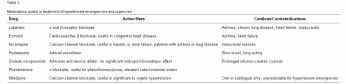
Diagnosis
While the patient remained in the ED, the nurse reported having difficulty using the automated machine to obtain blood pressures in the child’s upper extremities. Consequently, the nurse attempted to repeat the blood pressure in a lower extremity. At that time, the patient’s left lower extremity blood pressure was 115/51 mm Hg. Upon being notified of that value, blood pressure measurements in all 4 extremities were ordered by the ED physician, with the following results:
Right upper extremity: 180/100 mm Hg; left upper extremity: 180/88 mm Hg; right lower extremity: 121/78 mm Hg; left lower extremity: 123/74 mm Hg.
With the differential blood pressures, the ED physician made a presumptive diagnosis of coarctation of the aorta. An electrocardiogram and chest radiograph were obtained, and cardiology was consulted. The electrocardiogram showed normal sinus rhythm without axis deviation or hypertrophy. The chest radiograph showed right upper lobe airspace disease and a prominent heart silhouette.
Upon return from the radiology department, the patient was more awake and began complaining of abdominal pain. On reexamination, an abdominal bruit was heard. Echocardiography was ordered and performed by the on-duty technician. Preliminarily, the echocardiogram demonstrated only a mildly enlarged left ventricle and was otherwise unremarkable with no coarctation visualized.
As the echocardiography technician was leaving the ED, the emergency medicine resident caring for the patient asked if the whole aorta was visualized. The technician stated that by protocol, the echocardiography technicians will only scan to the level of the diaphragm. The resident asked the technician to take the ultrasound machine back into the patient’s room so that the resident could repeat the ultrasound examination independently. The resident was able to visualize the aorta and noted a narrowing in the upper abdomen, consistent with coarctation of the abdominal aorta. To confirm these findings, an abdominal CT with IV contrast was ordered. The CT revealed a long-segment (~3 cm) coarctation of the aorta at the level of renal artery suggestive of middle aortic syndrome.
Jeffrey R. Dingman MD and Pavan P. Zaveri MD
Division of Emergency Medicine, Children’s National Medical Center, Washington, DC
University of Maryland Medical Center, Baltimore, MD