The assessment of fecal incontinence in women
Fecal incontinence is a common problem in women after vaginal delivery. Overt sphincter damage from a third or fourth degree tear occurs in approximately 0.6% to 3% of women undergoing vaginal delivery. Twenty-nine percent to 48% of these patients develop anal incontinence between 3 months and 3 years after primary sphincter repair. Between 6.8% and 35% of primiparal and 12% and 44% of multiparal women have occult sphincter damage recognized on endoanal ultrasonography (EAUS)(Table 1). From one-third to two-thirds of women with such sphincter damage recognized on EAUS have bowel symptoms such as urgency or anal incontinenence.
After vaginal delivery pudendal nerve conduction can also be impaired. Prolonged pudendal nerve terminal motor latency (PNTML) is thought to occur in 42% of women who undergo vaginal delivery. Subsequent recovery is noted in 60% of these patients within the first 2 months after delivery.
Muscular and neurologic damage may also coexist. Indeed 60% of incontinent women who have sustained an obstetric injury to the external anal sphincter have prolonged PNTML.
Both severity and the prevalence of fecal incontinence increase with age, suggesting that other factors may also be involved. A progressive denervation of the anal sphincter muscles may be responsible for the delayed onset of fecal incontinence. But no clear correlation can be demonstrated between the weakness of the external anal sphincter and PNTML in elderly incontinent patients. These findings suggest that in this population a contribution to the weakness of external anal sphincter may be a decrease in the activity of the anterior horn cells in the spinal cord or the reduction in upper motor neurons caused by old age. The reduction in strength of the connective tissue fascia that occurs with the decline of estrogen production at menopause might weaken the pelvic floor and exacerbate any tendency to pudendal neuropathy caused by obstetric factors. This plethora of contributors mandates accurate assessment of anorectal and pelvic floor function in women with fecal incontinence to correctly identify the cause of the incontinence and to plan the most appropriate treatment.
Anorectal manometry
Anorectal manometry is a quantitative method of assessing resistance to spontaneous evacuation provided by the anorectal mechanism and the sensory capabilities of the rectum to provide a feeling of imminent emptying. It provides useful information in determining the causes of fecal incontinence and allows therapeutic decisions to be based on objective parameters. It also provides a baseline for comparison after treatment.
Anal canal pressures can be measured in a number of ways. The available methods use different sensory devices that include water-filled perfusion catheters, water- or air-filled balloons, sleeve catheters, and pressure transducers. Manometry can be performed with the stationary technique, the manual or stationary pull through technique, and the continuous pull through technique.
With the stationary technique the catheter is left in one position where the measurements are recorded. With the manual pull through technique the catheter tip is initially positioned 6 cm from the anal verge into the rectum and then gradually and progressively withdrawn at intervals of 1 cm. At each level the patient is asked to squeeze the sphincter muscles and hold for a few seconds and subsequently to strain to simulate evacuations while pressures are recorded. A 20- to 30-second interval is given between measurements to stabilize the anal pressure. Using multilumen catheters, the continuous pull through technique can assess anal canal pressures on a cross-sectional basis at each recording station from the anal verge, allowing examination of pressures in each quadrant of the anal canal. This measurement provides radial and longitudinal pressure profiles from which vector diagrams, when plotted together, can generate a three-dimensional picture of the anal canal, known as the vector volume; the vector volume analysis demonstrates the anal canal pressure symmetry.
Several methods of analysis have been introduced in an attempt to quantify and express anal canal manometric asymmetry. The total radial asymmetry measures the mean asymmetry of the anal canal from the sum of the deviance of minimum vector volumes from the mean of all vector volumes at each level within the anal canal. The vector symmetry index (VSI) is calculated dividing the anal canal into eight segments, the volume of each being the sum of the vector volumes within each segment. Reconstruction of these manometric data generates a three-dimensional wire frame and the VSI represents the ratio of minimum segment volume to the maximum segment volume. A VSI of 1.0 represents perfect symmetry; varying degrees of asymmetry lie between 0 and 1.0. Physiologic radial asymmetry of the anal canal has been demonstrated with anterior quadrant pressures being lower than those laterally and posteriorly. Nevertheless, symmetry of the anal canal pressures can be distorted or reduced in incontinent patients, so they have been used as parameters for evaluating anorectal pathology. But it has been demonstrated that vector volume analysis shows a lower squeeze pressure profile than does the stationary pull through technique. This difference might be from the stabilization period that is obtained at each single point of the anal canal with the stationary pull through technique, or perhaps it reflects the patent’s inability to perform sustained squeeze during the catheter withdrawal process. So the vector volume manometry theoretically provides a less reliable pressure measurement than does the stationary pull through technique.
Only 13.2% of patients with isolated decreased electromyographic activity have a sphincter defect localized by vector volume analysis and only 11% of patients with a sphincter defect demonstrated on EAUS have identical findings on vector volume analysis. Both standard and vector manometry findings have shown no correlation with morphologic parameters as demonstrated on EAUS and endoanal magnetic resonance imaging (MRI). But a more recent study found a good correlation among anal manometry parameters, VSI, EAUS, and clinical findings. Total asymmetry failed to show this correlation. It was concluded in this study that anal vector manometry using VSI analysis of anal squeeze pressure with its high sensitivity is complementary to EAUS because it provides information regarding anal sphincter function and verification of anal sphincter disruption.
Resting pressure is determined mostly by the internal anal sphincter, and the external sphincter is mainly responsible for squeeze pressure. When normal subjects contract their anal sphincters, the anal canal pressure rises between 150% and 250%. Resting is slightly lower in women than in men; and squeeze pressure is significantly lower in women, anal canal pressures also reduce with age. Squeeze pressure is more affected than is resting pressure with age, and the decrease is more marked in women than in men.
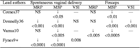
Resting and squeeze pressures are both decreased after vaginal delivery. Squeeze pressure is more frequently reduced and is more pronounced after forceps delivery (Table 2). Resting pressure is also reduced after proctectomy with coloanal anastomosis and by pelvic irradiation.
Table 2. Postdelivery Manometric Changes in Primiparal Women
Anal canal length can also be manometrically assessed with stationary pull through technique. Anal canal length is defined as the length of the anal canal at which resting pressures equal or exceed 20 mmHg in all four quadrants. The high pressure zone is identified from distal to proximal and is represented by the zone in the anal canal in which pressures are greater than 50% of the average resting pressure. Length of the anal canal and the high pressure zone are both shorter in women than in men.
A sensation of rectal filling is essential for normal defecation and plays an important role in the continence mechanism. Rectal sensation can be tested by balloon distension of the rectum. A balloon connected to a fine catheter is inserted into the rectum lying, once inflated, on the pelvic floor. The balloon can be distended either by air or water. Three measurements are taken. The rectal sensory threshold corresponds to the minimum volume felt in the rectum; the sensation of fullness is the maximal volume injected into the rectum before there is an urgent desire to defecate; and the maximum tolerable volume is the volume causing unbearable discomfort.
Rectal capacity is calculated by subtracting the first appreciation of rectal filling (sensory threshold) to the volume producing the urge to evacuate (sensation of fullness); so rectal capacity determines the frequency of evacuation. Rectal compliance is defined by ratio of rectal capacity to gradient pressure and is responsible for the degree of urgency of evacuation. Because maximum tolerable volume is very uncomfortable and does not add any clinically relevant information, it is not routinely measured. Many factors can affect rectal capacity and compliance, both of which are reduced in patients with chronic inflammatory disease, radiation proctitis, and rectal prolapse.
The rectoanal inhibitory (RAIR) or sampling reflex is a transient rise followed by a profound fall in resting anal canal pressures after distention of the rectum with small volumes of air or liquid; the amplitude and duration of the reflex increase with larger distending volumes. The RAIR can be assessed by placing a soft balloon in the rectum and recording the anal canal pressure at the site of maximal basal pressure. The reflex is important for initiating and controlling evacuation because relaxation of the internal sphincter allows the fecal bolus to contact the sensitive epithelium of the mid-anal canal (anal transition zone). Once the bolus is recognized the external sphincter and puborectalis either relax, allowing normal evacuation or, if it is socially unacceptable, the pelvic floor contracts, forcing fecal material back in to the rectum from the anal canal, which returns to its normal position. The RAIR is absent in patients with Hirschsprung’s and Chagas’ disease, but is preserved in patients with high cord compression, spinal anesthesia, or pudendal nerve block, after administration of muscle relaxant and after resection of the rectum if the anal canal is preserved, suggesting that intrinsic myenteric nerve plexuses play a major role. The RAIR is also absent in patients with rectal prolapse and abnormal in patients with scleroderma and other connective tissue disorders. In incontinent patients RAIR may be absent or more attenuated and the threshold higher.
The anocutaneous reflex is assessed by stroking the perianal skin and recording, with the catheter stationary in the high pressure area of the anal canal, the external sphincter contraction that follows. Absence of the anocutaneous reflex confirms denervation of the external sphincter suggested by an absent voluntary squeeze. Additional confirmation is given by the absence of reflex involuntary contraction while coughing and during the Valsalva maneuver. But the anocutaneous reflex is absent in some incontinent patients.
Ambulatory manometry
Coordination of pressures and motor events between the rectum and anal canal is important to maintain an efficient mechanism of continence. Ambulatory manometry assesses the sigmoidal, rectal, and anal motor activity continuously over a period of 24 hours. This allows recordings when the subject is ambulatory and when asleep. The technique involves the introduction into the rectum of a flexible microtransducer catheter with a central lumen for insertion over a wire. The catheter has six sensors positioned at different levels along the catheter from 2 to 25 cm from the tip. When the catheter is in place, the sensors will be distributed at different heights from the anal canal to the lower sigmoid colon, with the two lowest ones positioned in the anal canal. The catheter is then connected by a cable to a small portable battery-powered recorder. At the end of the examination, data are downloaded into a personal computer for analysis. This technique provides a monitoring of the temporal relationships between rectal and anal canal motor events that is not obtained with conventional manometry. So ambulatory manometry offers a better correlation with the cause of functional anorectal disorders than does conventional manometry.
Endoanal ultrasonography
EAUS provides a morphologic assessment of the internal and external anal sphincters, the puborectalis muscle, and the rectovaginal septum. The examination is undertaken with a rotating endoprobe with a 7- or 10-mm MHz transducer that allows 360-degree evaluation of the anal canal. The use of a higher frequency transducer has the benefit of improved tissue resolution and detail in the near field. The anal canal is shown in alternate different layers of hypo-, hyper-, or mixed echogenicity that allows a clear identification of the internal and external anal sphincters. The examination does not require any preparation or sedation and is normally well tolerated by the patient.
EAUS has proved to be at least as good as electromyography (EMG) in assessing patients with fecal incontinence. It provides information about the integrity of the internal and external anal sphincters and detects sphincteric defects with an accuracy of 90% to 100%. When directly compared to clinical examination, manometry, and EMG, EAUS was the only test to achieve 100% of sensitivity, predictive value, and accuracy. So EAUS is a very valuable tool in planning restorative surgery. It also is an effective technique in assessing results after sphincter repair.
Persistent incontinence after sphincter repair is most frequently from a disruption of the repair. Because EAUS is a simple and well-tolerated procedure it has now, in many centers, largely replaced the more invasive technique of EMG. But EMG may detect functional abnormalities or even absence in incontinent patients with normal anal sphincters on EAUS. So the two techniques are complementary and not mutually exclusive.
Electromyography
EMG provides an assessment of the neuromuscular integrity of the anal canal by recording the motor unit action potentials generated by the external sphincter and puborectalis muscle fibers at rest, and during voluntary contraction during evacuation and in response to various reflexes. Although continuous electrical activity can be recorded by the anal EMG even at rest, EMG activity of pelvic floor muscles increases with rectal or bladder distention, change of body position, and during reflexes such as cough, anal scratch, and bulbocavernous stimulation. Electromyographic activity normally ceases during relaxation of striated sphincter muscles as seen during evacuation. The main purposes of anal EMG are to assess the functional activity of the anal sphincter and pelvic floor muscles and assess the presence of neurologic damage. But, anal EMG is not a useful test to differentiate the abnormal electrical activity of myopathic and neurogenic disorders.
Electrical activity can be recorded in different ways. Concentric needle electrodes consist of a bare-tipped 0.1-mm diameter still wire with an insulating resin. The needle is inserted lateral to the anal canal into the external sphincter or puborectalis muscle. The insertion is guided by the examiner’s contralateral index finger in the anal canal. The electrical activity is recorded from each of the four quadrants of the external anal sphincter. Monopolar wire electrodes were developed to prevent the possibility of sliding and to allay the fear of pain caused by the rigid concentric needle. The thin Teflon-coated monopolar wire electrode is removed 4 mm from the tip. It is inserted by guidance of a lumbar puncture needle and kept in position by a small hook at the tip.
Single fiber EMG electrodes are 25-μm electrodes filled with resin that can record the activity of individual muscle fibers within a motor unit. The single fiber EMG allows calculation of the fiber density and neuromuscular jitter. The fiber density consists of the mean number of single fiber action potential in 20 different positions within the detected muscle. The neuromuscular jitter is the time interval between two action potentials in the same motor unit recorded in the uptake area of a single fiber EMG, and is a measure of end-plate function. The fiber density is increased in case of denervation during the reinervation process, which results in more muscle fibers inervated by an individual axon (fiber-type grouping). Jitter is increased in neurologic disorders, such as myasthenia gravis, and where there is peripheral nerve sprouting in reinervation. Newly formed sprouts are not initially myelinated and do not transmit nerve impulses well.
Cutaneous surface electrodes placed in pairs on the perianal skin to obtain the electrical activity of the external anal sphincter muscle were developed because of the discomfort associated with needle electrodes. But recording was inaccurate and interpretation subjective because of the interference from the electrical activity of adjacent large muscles such as the adductor and gluteus. A disposable anal plug electrode consisting of two longitudinal or circular silver wires mounted on the surface of a trochlear-shaped sponge or a plastic plug was developed. The longitudinal electrodes were found to have a better correlation with fine wire electrodes than the circular ones.
Anal plug EMG is less invasive than wire electrodes, and EMG has now gained widespread clinical use for biofeedback therapy. In a recent study, a new quantitative surface EMG signal analysis showed a significant positive correlation with the single fiber EMG, suggesting the possibility of becoming a new powerful noninvasive means of investigation for patients with pelvic floor disorders.
Abnormal EMG findings can result from underlying conditions. In case of muscular damage there are markedly decreased motor unit potentials at rest, during squeeze, reflex elicitation, and evacuation. Neurogenic damage, when partial, shows a fiber-type grouping (reinervation) appearance with increased amplitude, prolonged duration, polyphasic motor unit potentials, and an increased neuromuscular jitter between individual components. In case of complete denervation there are markedly decreased or absent motor unit potentials. Functional disorders such as anismus show no change or even increased motor unit potentials during evacuation.
Although EAUS has largely replaced EMG in the assessment of the anal sphincter in patients with fecal incontinence, it only provides anatomic details regarding the sphincter muscles, but does not offer any information regarding their neuromuscular integrity. Conversely, EMG assesses function. The analogy to gross and microscopic anatomy may be applicable. EAUS analyzes only structure, and EMG allows evaluation of function.
Pudendal nerve terminal motor latency
PNTML measures the length of time required for a fixed electrical stimulus to travel along the pudendal nerve between the ipsilateral ischial spine and the anal verge. PNTML provides evaluation of pelvic floor neuromuscular integrity. Both pudendal nerves are transanally stimulated as they traverse the ischial spine, with a dedicated electrode (St Mark’s Pudendal Electrode 13L40, Dantec Electronics, Bristol, UK) placed on the tip of the examining finger and connected to a pulsed stimulus generator. The electromechanical response of the external anal sphincter is received by the electrode placed at the base of the examining finger and registered by an oscilloscope. Analyzing the elapsed time from the beginning of the stimulus to the moment of the muscle contraction allows the speed of contraction to be calculated. Each of the two nerves is stimulated three times and the average latency recorded; different factors seem to affect PNTML.
Several studies have suggested that pudendal neuropathy is an age-related phenomenon. In addition, chronic straining followed by perineal descent has been anecdotally postulated to cause a stretch injury to the pudendal nerves and prolong the PNTML. But several large studies demonstrated no association between perineal descent and pudendal neuropathy, either in patients with chronic constipation or fecal incontinence. Evidence that association of perineal descent and pudendal neuropathy may be a coincidental phenomenon has been accumulated and pudendal neuropathy appears to relate to age more than any other variable.
Vaginal delivery also affects PNMTL, the risk of pudendal nerve damage increases with multiparity, forceps delivery, increased duration of the second stage of labor, third degree perineal tear, and high birth weight. Damage to the pudendal nerve occurs in 16% of primiparal women and 15% of multiparal women, and up to 60% of women with obstetric external anal sphincter injury have a pudendal neuropathy, subsequent recovery occurs in approximately 15% of these injured patients.
It has been suggested that fecal incontinence after vaginal delivery can result from damage to the innervation of the pelvic floor muscle. In addition, an important association between abnormal latency and the development of a sphincter defect in primiparal women has been found. Regardless of whether pudendal nerve damage is responsible for symptoms of fecal incontinence after vaginal delivery, such damage can be a factor predictive of outcomes after anterior overlapping sphincteroplasty. But not all the series have confirmed these data (Table 3)
Table 3. Pudendal Neuropathy and Surgical Outcomes
Cinedefecography
Cinedefecography is a radiographic method of evaluating the dynamics of evacuation. This technique involves the filling of the rectum with 50 mL of liquid barium, followed by the introduction of 100 to 200 mL of barium paste with a consistency similar to stool. The patient is seated on a translucent commode and the anorectum laterally visualized by fluoroscopic technique. Lateral films are obtained at rest, and during squeeze and maximum strain after which the mechanism of evacuation is recorded by video. Parameters evaluated during resting, squeeze, and straining phases include the anorectal angle, puborectalis length, and perineal descent. The changes in these parameters during the different phases are also analyzed. The examination is not routinely indicated in patients with incontinence because these patients often cannot retain the contrast, and it seldom contributes to the diagnosis in these patients. But in selected cases it provides information about the presence of associated factors such as intussusception, rectal prolapse, and increased perineal descent.
Magnetic resonance imaging
In general MRI has a high sensitivity to soft tissue abnormalities, but conventional MRI with external body coil does not provide adequate definition of the anal sphincters. Dedicated endoanal coils have been developed to increase the resolution in the assessment of the anal canal. Endoluminal MRI, with its high intrinsic contrast resolution and the high spatial resolution of an endoanal coil, has provided detailed definition of the anal canal anatomy. The examination is normally undertaken with the patient in the supine position with a cylindrical rigid receiver coil placed in the anal canal. The investigation does not require any preparation and, except in patients with extensive perianal fistula or anal stenosis, is well tolerated by the patients. With endoluminal MRI, all layers of the anal sphincter can be easily identified. Lesions of the external anal sphincter such as sphincter defects and scar tissue are demonstrated with an accuracy of 90% to 95%.
A comparative study of endoluminal ultrasonography and endoluminal MRI in 22 patients with fecal incontinence has demonstrated that endoluminal MRI is superior to endoanal utrasonography in detecting defects of the external anal sphincter (accuracy = 91% versus 73%). In that study, EAUS was performed with a 7-MHz transducer. It is likely that with the use of a near-field focusing 10-MHz transducer, the definition and accuracy of the investigation will increase. Conversely, it seems that the internal anal sphincter, which is well seen by EAUS, is more difficult to define by endoanal MRI. A prospective study of 52 consecutive unselected patients with anal incontinence found that 12 errors related to internal sphincter integrity were made at endoanal MRI compared with only one at EAUS. The same study found endoanal MRI and EAUS equivalent in diagnosing external anal sphincter injury. Although endoluminal MRI can easily identify atrophy of the external anal sphincter, as yet there are no EAUS criteria for external sphincter atrophy. External sphincter atrophy, as identified on endoanal MRI, might also represent a predictor of the negative outcomes of anterior anal repair.
Anal sensation
The sensory function of the anorectum is involved in discrimination between flatus, liquid, and solid stool. It is unclear whether the integrity of sensory function in the anal canal plays a role in the maintenance of continence. But it is likely that minor degrees of sensory impairment are not by themselves causative of incontinence in patients with otherwise normal anorectal function. In patients with poor sphincter function though, the sensory abnormality might make patients unaware of the seepage of fecal material through the anal canal and determine a worsening of symptoms.
Anal sensation was originally measured using pinprick and metal plates at different temperatures. Such techniques are crude and provide only qualitative data. Mucosal electrosensitivity and thermal sensitivity are the two currently used tests to assess anal sensation. The mucosal electrosensitivity technique requires a current generator connected to a probe with the active electrode and a reference electrode. The reference electrode is placed on the patient’s thigh, while the probe is lubricated with a conductive gel and introduced into the anal canal so that the electrode lies in the upper anal canal. The current is then increased in 1-mA increments until the subject feels a tingling or prickling sensation. The voltage at which sensation is first felt is recorded. The procedure is repeated three to five times at the same level and the mean of these readings is documented as the threshold of sensation for that particular level. The probe is withdrawn and the same process repeated for the mid- and lower anal canal.
Thermal sensitivity assesses the thermal threshold of the anal canal. A 1-cm probe introduced into the anal canal through which water of different temperature can be pumped provides cold and hot stimuli. The temperature of the thermode can be maintained at 37 °C and then rapidly increased or decreased by 4.5 °C. A small thermocouple placed on the outside of the probe continuously records the temperature of the thermode-mucosal interface. The thermal threshold is assessed for four temperature ranges: normal to hot, hot to normal, normal to cold, and cold to normal. The assessment is performed in the upper, mid-, and lower anal canal and a median value of the four thresholds represents the minimum detectable temperature change for that level. The anal canal is very sensitive to thermal stimuli and this sensitivity is diminished in patients with idiopathic fecal incontinence.
In conclusion, fecal incontinence is a complex problem, often of multifactorial origin. Although the use of all tests currently available for assessment of this condition is generally not required, complete functional and anatomic assessments of the anorectum, anal sphincters, and pelvic floor are mandatory in all women with fecal incontinence to correctly identify the cause and type of incontinence and allow correct treatment. Determining the most appropriate tests will largely depend on the patient’s history and symptoms and can vary for each patient.
References
1. A.H. Sultan, M.A. Kamm, C.N. Hudson et al., Anal sphincter disruption during vaginal delivery. N Engl J Med 329 (1993), pp. 1905–1911.
2. A.H. Sultan, M.A. Kamm, C.N. Hudson and C.I. Bartram, Third degree obstetric anal sphincter tears: risk factors and outcome of primary repair. BMJ 308 (1994), pp. 887–891.
3. K. Haadem, S. Ohrlander and G. Lingman, Long-term ailments due to anal sphincter rupture caused by delivery - a hidden problem. Eur J Obstet Gynecol Reprod Biol 27 (1988), pp. 27–32.
4. C.J. Walsh, E.F. Mooney, G.J. Upton and R.W. Motson, Incidence of third-degree perineal tears in labour and outcome after primary repair. Br J Surg 83 (1996), pp. 218–221.
5. K. Haadem, J.A. Dahlstrom and L. Lennart, Anal sphincter competence in healthy women: clinical implications of age and other factors. Obstet Gynaecol 78 (1991), pp. 823–827.
6. S.M. Sorensen, H. Bondesen, O. Istre and P. Vilmann, Perineal rupture following vaginal delivery. Acta Obstet Gynecol Scand 67 (1988), pp. 315–318.
7. M.B. Neilsen, C. Hauge, O.O. Rasmussen et al., Anal endosonographic findings in the follow-up of primarily sutured sphincteric rupture. Br J Surg 79 (1992), pp. 104–106.
8. J. Zetterstrom, A. Mellgren, L.L. Jensen et al., Effect of delivery on anal sphincter morphology and function. Dis Colon Rectum 42 (1999), pp. 1253–1260.
9. M. Fynes, V. Donnelly, M Behan et al., Effect of second vaginal delivery, on anorectal physiology and faecal continence: a prospective study. Lancet 353 (1999), pp. 983–986.
10. A. Varma, J. Gunn, A. Gardiner et al., Obstetric anal sphincter injury. Prospective evaluation of incidence. Dis Colon Rectum 42 (1999), pp. 1537–1543.
11. S.J. Snooks, M. Setchell, M. Swash and M.M. Henry, Injury of innervation of pelvic floor sphincter musculature in childbirth. Lancet 2 (1984), pp. 546–550.
12. S.J. Snooks, M,M. Henry and M. Swash, Faecal incontinence due to external sphincter division in childbirth is associated with damage to the innervation of the pelvic floor musculature. Br J Obstet Gynaecol 92 (1985), pp. 824–828.
13. A.J. Campbell, J. Reinken and L. McCosh, Incontinence in the elderly: prevalence and prognosis. Age Ageing 14 (1985), pp. 65–70.
14. S. Laurberg and M. Swash, Effect of aging on the anorectal sphincters and their innervation. Dis Colon Rectum 32 (1989), pp. 737–742.
15. S.J. Snooks, M. Swash, S.E. Mathers and M.M. Henry, Effect of vaginal delivery on the pelvic floor: a 5-year follow-up. Br J Surg 77 (1990), pp. 1358–1360.
16. F. Beersiek, A.G. Parks and M. Swash, Pathogenesis of anorectal incontinence: histometric study of the anal sphincter musculature. J Neurol Sci 42 (1979), pp. 111–127.
17. I.J. Dorfman and T.M. Bosley, Age related changes in peripheral and central nerve conduction in man. Neurology 29 (1979), pp. 38–44.
Presented at the American College of Surgeons 86th Annual Clinical Congress, Chicago, IL, October 2000. Presentation sponsored by the Advisory Council for Gynecology and Obstetrics and the Advisory Council for Colon and Rectal Surgery.
Pasquale Giordano MD and Steven D. Wexner MD, FACS, FRCS, FRCS(ED)
Department of Colorectal Surgery, Cleveland Clinic Florida, Weston, FL USA