
Clinical Examination of the Anorectum
Key points
- An accurate examination of the anorectum is required in the setting of symptoms referred to that region and should be part of a patient's routine physical examination (1).
- The next component of the examination involves palpation whereby the gloved and lubricated index finger is used to identify tenderness, mass lesions, and areas of induration and to examine the sacrococcygeal, perineal, and perianal tissues (2).
- A fistula tract may be traced and pus may be expressed from the secondary opening.
- Attention is then focused on the posterior rectal wall above the anorectal ring to identify palpable rectal masses, ulcerated tumors, pedunculated polyps, and abscesses or submucosal rectal masses.
An accurate examination of the anorectum is required in the setting of symptoms referred to that region and should be part of a patient's routine physical examination (1). Care should be taken to maintain the patient's dignity and to minimize any embarrassment. The rectal examination is performed with the patient lying in the left lateral decubitus position with knees bent toward the chest. Proper draping is required to expose only the perineum, and good lighting is essential for a careful inspection to exclude obvious abnormalities of the anal and perianal area. Inspection is performed with proper retraction of the buttocks. The buttocks, sacrococcygeal region, vulva or base of the scrotum, and upper thighs should be examined first and then the anal and perianal area.
The next component of the examination involves palpation whereby the gloved and lubricated index finger is used to identify tenderness, mass lesions, and areas of induration and to examine the sacrococcygeal, perineal, and perianal tissues (2).
A fistula tract may be traced and pus may be expressed from the secondary opening. The gloved finger is placed at the anal orifice and gradually inserted into the anal canal where the intersphincteric groove is palpable circumferentially and abscesses or tenderness can be identified. Internal hemorrhoids are often not palpable unless they have formed a thrombosis.
Attention is then focused on the posterior rectal wall above the anorectal ring to identify palpable rectal masses, ulcerated tumors, pedunculated polyps, and abscesses or submucosal rectal masses. Stool in the rectum is deformable but masses are not—they are often fixed, superficially mobile, or tethered. The anorectal ring is prominent in the lateral and posterior region; after the patient is instructed to contract the rectal muscle, the strength of contraction is noted to identify any underlying sphincter injuries.
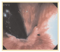
To examine the anal canal or mucosa, anoscopy or flexible sigmoidoscopy is sometimes used. The anoscope is about 7 cm long and 2 cm in diameter with a beveled tip. After insertion into the anal canal, it is withdrawn to the dentate line where the papillae and crypts are inspected and fistula openings and fissures can be identified. The anoscope should not be used as the sole instrument of inspection if there is rectal bleeding. Flexible sigmoidoscopy and colonoscopy performed by a qualified endoscopist can be used to examine the rectum and anorectum on withdrawal and retroflexion (Figure 1).
---
Figure 1. The demarcation of "white" squamous cell mucosa and the "pink" columnar mucosa is the dentate line (denoted by the arrow).---
Written and revised by
Andrey Nailor, M.D.
Our doctors write and review articles for accuracy and keep them up to date.
health.am provides general information, not medical advice. Always consult a qualified health professional about your own health.
Explore health topics
More in this topic
Digestive Health → Guide
Treatment Strategy
The natural history of Crohn's disease involves fiuctuating periods of disease quiescence punctuated by episodes of exacerbation. Cumulative bowel injury occurs over time with chronic smoldering inflammation and is…
25 August 2015
GuideMethotrexate
Methotrexate is a structural analogue of folic acid that competitively inhibits its binding to its receptors and has demonstrated effectiveness for treating several autoimmune diseases, including rheumatoid arthritis…
25 August 2015
GuideThe Story Behind Crohn and Ginzburg and Oppenheimer(CGO) Collaboration
Crohn, Ginzburg, and Oppenheimer all graduated from Columbia University's College of Physicians and Surgeons and subsequently succeeded in becoming house staff at the Mount Sinai Hospital. Their careers were certainly…
25 August 2015