Vulvar Atypias - Cancer of the Vulva
Key points
- There has been a lack of uniformity in defining vulvar atypias.
- There are minimal data to support vulvar dystrophies being a cause of cancer.
- The International Society for the Study of Vulvar Disease has provided a standard nomenclature for these lesions, and this has been adopted by the International Society for Gynecologic Pathology (Table 100-1).
- In 1985, the nomenclature was replaced by the terms carcinoma in situ or vulvar intraepithelial neoplasia (VIN).
VULVAR ATYPIAS
There has been a lack of uniformity in defining vulvar atypias. There are minimal data to support vulvar dystrophies being a cause of cancer. The International Society for the Study of Vulvar Disease has provided a standard nomenclature for these lesions, and this has been adopted by the International Society for Gynecologic Pathology (Table 100-1). In 1985, the nomenclature was replaced by the terms carcinoma in situ or vulvar intraepithelial neoplasia (VIN). In addition, the nonpremalignant squamous changes previously termed hyperplastic dystrophy are now considered under the classification of squamous hyperplasia.
Vulvar atypias can present with a variety of symptoms. The most common is irritation or itching; however, 20% of patients are asymptomatic.
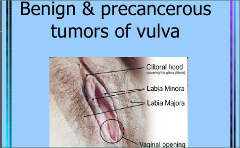
Grossly, the lesions can be flat, raised (maculopapular), or verrucous. In color, they may be brown (hyperpigmented), red (erythroplastic), white, or discolored.
White lesions can appear to have a whitish, thickened keratin layer (leukoplakia) or a diffuse, white, brittle, paperlike appearance (lichen sclerosus) (Figure 100-2). Areas of squamous hyperplasia (hyperplastic dystrophy) and dysplasia can also have a white appearance. Unlike lichen sclerosus, however, the tissue often is thickened, and the process tends to be focal or multifocal rather than diffuse.
Microscopically, atypical changes in the vulvar epithelium consistent with preinvasive lesions usually are marked by loss of maturation of the squamous epithelium. There is increased mitotic activity and an increase in the nuclear cytoplasmic ratio. Mild dysplasia (VIN I) is diagnosed if changes involve the lower third of the epithelium. If there is moderate dysplasia (VIN II), then half to two-thirds of the epithelium is affected. If there is severe dysplasia (VIN III), then two-thirds of the epithelium is affected. For carcinoma in situ (also classified as VIN III) of the vulva, the full thickness of the epithelium usually is abnormal.
It often is difficult to distinguish between benign squamous hyperplasia and intraepithelial neoplasia. Crum and colleagues suggested that nuclear size may be helpful in diagnosis because intraepithelial neoplasia of the vulva almost always contains nuclei that are fourfold or greater in size compared with benign condyloma or non-neoplastic epithelium.
The best method of establishing a diagnosis is a high index of suspicion and early biopsy.
Several methods also can be used to help assess these lesions. Cytology, colposcopy, and toluidine blue O can be used cautiously before biopsy. In general, however, cytologic evaluation of the vulva has not been helpful as a screening examination because the vulvar skin often is thickened and keratinized. Colposcopic examination of the vulva is difficult because unlike cervical lesions, the changes are difficult to recognize. Therefore, colposcopic examination is not used for routine vulvar examination; rather, it is primarily employed for patients who are being evaluated or followed for vulvar atypia or intraepithelial malignancies. The toluidine blue O test is nonspecific and stains nuclei in the superficial part of the epithelium. Colposcopy is performed after applying a 1% aqueous solution of toluidine blue O to the vulva for 1 minute and decolorizing the tissue with 1% acetic acid. Areas that retain the stain are biopsied. A positive test, however, does not always indicate a premalignant condition because 20% of benign areas on the vulva stain positively.
Written and revised by
Gerald Hurowitz, M.D.
Our doctors write and review articles for accuracy and keep them up to date.
health.am provides general information, not medical advice. Always consult a qualified health professional about your own health.
Explore health topics
More in this topic
Cancer → News
Study may help reassure women taking tamoxifen for breast cancer
A study led by Loyola Medicine researchers may help reassure patients who worry the breast cancer drug tamoxifen could increase their risk of uterine cancer. The multi-center study was presented during the 2016 annual…
7 June 2016
 News
High fruit intake during adolescence linked with lower breast cancer risk
Two linked papers in The BMJ this week shed new light on the relation of alcohol and diet with breast cancer and heart disease . The first study reports that high fruit consumption during adolescence may be associated…
13 May 2016
 News
Andrei Goga's UCSF team finds new approaches to eradicate aggressive breast cancers
Conventional chemotherapy generally fails to eradicate aggressive breast cancers due to the early distant metastasis that can occur in these diseases. Triple-negative breast cancer (TNBC) is a particularly aggressive…