Invasive Vulvar Carcinomas
Key points
- The International Federation of Gynecology and Obstetrics (FIGO) has defined four clinical stages of vulvar carcinoma.
- There is no uniformly accepted definition regarding microinvasive carcinoma of the vulva, and this may result from confusion in measuring the depth of invasion.
- The prognosis of a patient with vulvar carcinoma relates to the stage of disease (Figure 100-4) and to the nodal status.
- In addition, many centers use the tumor, node, metastasis (TNM) classification.
INVASIVE VULVAR CARCINOMAS
The International Federation of Gynecology and Obstetrics (FIGO) has defined four clinical stages of vulvar carcinoma. In addition, many centers use the tumor, node, metastasis (TNM) classification. In 1988, the FIGO staging was modified to reflect lymph node status and the location of the tumor (Table 100-2). In this classification, a tumor that is located on the perineum is no longer considered to be stage III. In 1995, FIGO instituted a subclassification of stage I into IA and IB, based on whether there is stromal invasion greater than 1 mm.
Squamous Cell Carcinoma Squamous cell carcinomas comprise approximately 90% of primary vulvar malignancies. Grossly, these carcinomas usually appear as ulcerated or polyploid masses on the vulva. Biopsy reveals the characteristic histologic appearance: the tumor appears in nests and cords of squamous cells infiltrating the stroma, often with islands of keratin.
The pattern of spread for this carcinoma relates to the intricate lymphatic drainage of the vulva (Figure 100-3). Tumors located in the middle of either labium initially drain to the ipsilateral inguinal femoral nodes, whereas midline perineal tumors can spread either to the left or right side. Using technetium 99m colloid, Iversen and Aas showed that when radioactivity was injected to one side of the vulva, 98% of it localized in the ipsilateral nodes and less than 2% in the contralateral nodes. Tumors along the midline in the clitoral or urethral areas may spread to either groin. From the inguinal-femoral nodes, lymphatic spread continues to the deep pelvic iliac and obturator nodes. Although there has been concern in the past that tumors in the clitoral-urethral area could spread directly to the deep pelvic nodes, current evidence indicates that this is rare.
Stage I Microinvasive Carcinoma
There is no uniformly accepted definition regarding microinvasive carcinoma of the vulva, and this may result from confusion in measuring the depth of invasion. Microinvasion has been defined by some as a small lesion of the vulva less than 2 cm in diameter and invading less than 3 mm. The International Society for the Study of Vulvar Diseases has recommended that the term microinvasive be dropped and that stage IA be used for tumors less than 2 cm in diameter and invading less than 1 mm from the epidermal-stromal junction, the basement membrane.
{embed="cr/stat-menu-vulva"}
Different clinical results have been reported with this definition. Spread to regional lymph nodes has varied from 0 to 10% in tumors with less than a 5 mm depth of invasion. For example, Hoffman and colleagues reported no nodal metastases in 43 patients whose tumor invaded less than 2 mm. Lesions that were at risk of spreading to inguinal nodes included tumors with confluent tongues rather than those with individual tongues merely extending into the stroma. Hacker and colleagues, however, reported that six of seven patients with invasion of less than 3 mm had regional node involvement.
Written and revised by
David Cohen, M.D.
Our doctors write and review articles for accuracy and keep them up to date.
health.am provides general information, not medical advice. Always consult a qualified health professional about your own health.
Explore health topics
More in this topic
Cancer → News
Non-invasive imaging can detect mutations within a brain tumor
Researchers at Winship Cancer Institute have developed a technique for detecting an "oncometabolite", a chemical produced by some brain tumors’ warped metabolism, via non-invasive imaging. Their approach could allow…
23 March 2012
 News
MRI May Be Noninvasive Method to Measure Breast Cancer Prognosis
Quantitative magnetic resonance imaging measures were associated with prognostic tumor markers, demonstrating the potential of magnetic resonance imaging for prediction of disease prognosis and stratification of…
9 December 2011
 Overview
Vulvar Cancer
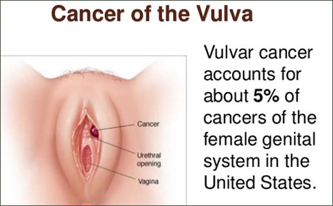
Vulvar cancer is an uncommon malignancy, responsible for only 5% of all gynecologic neoplasms , but its incidence is on the rise.
5 December 2011