The Mammogram Report: The Breast Imaging Reporting and Data System (BI-RADS™) Lexicon
Gerald Hurowitz, M.D.·Published 14 June 2008·7 min read
Key points
The Breast Imaging Reporting and Data System (BI-RADS™) lexicon was developed by the American College of Radiology (ACR) in an effort to standardize terminology used in reporting mammograms.
Benign Finding: This is also a negative mammogram, but the interpreter may wish to describe a finding.
Probably Benign Finding - Short Interval Follow-up Suggested: A finding placed in this category should have a very high probability of being benign.
Suspicious Abnormality - Biopsy Should Be Considered: These are lesions that do not have the characteristic morphologies of breast cance but have a definite probability of being malignant.
The Breast Imaging Reporting and Data System (BI-RADS™) lexicon was developed by the American College of Radiology (ACR) in an effort to standardize terminology used in reporting mammograms.
The lexicon includes specific terminology for reporting breast composition, mammographic findings, such as masses and calcifications, and overall assessment of the likelihood of malignancy.
Breast Composition Breast composition may be one of four patterns of increasing density:
almost entirely fat
scattered fibroglandular densities
heterogeneously dense
extremely dense
The higher the breast density, the lower the sensitivity of mammography.
Masses A mass is defined as a space-occupying lesion seen in two different projections. If a possible mass is seen on only one view, it is called a density until its three-dimensionality is confirmed. The lexicon includes specific terms to describe masses and calcifications. Possible mass shapes are round, oval, lobular or irregular. Architectural distortion is a special case of shape, in which there are radiating spicules with no central mass visible.
Mass margins can be circumscribed (well defined or sharply defined), microlobulated (undulant with short cycles), obscured (hidden by adjacent fibroglandular tissue), indistinct (ill defined) or spiculated (with lines radiating from the margins of the mass, which suggests a lesion that is invading surrounding tissue or inciting desmoplastic reaction). Special cases of masses include solitary dilated ducts (usually of minor significance), intramammary lymph nodes (of a reniform shape, with radiolucent notch due to fat in the hilum), asymmetric breast tissue (may be important when it corresponds to a palpable asymmetry), and focal asymmetric density (visible as asymmetry with similar shape on two views, but lacks the borders and conspicuity of a true mass).
Density of masses can be high, equal (isodense), low, or fat-containing/radiolucent (e.g., oil cyst, lipoma, galactocele or mixed lesions such as hamartoma).
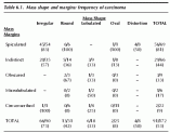
The frequency of carcinoma as a function of mass shape and margins in a study by Liberman et al of lesions that had surgical biopsy is demonstrated in Table 6.1. The highest frequency of carcinoma was observed in masses that had irregular shape or spiculated borders. {pagebreak}
Calcifications The BI-RADS™ lexicon describes calcification morphology (shape) and distribution. Calcification morphology is divided into three groups: typically benign, intermediate level of concern and higher probability of malignancy.
Written and revised by
Gerald Hurowitz, M.D.
Our doctors write and review articles for accuracy and keep them up to date.
health.am provides general information, not medical advice. Always consult a qualified health professional about your own health.
Typically benign morphologies include skin (lucent centered), vascular (parallel tracks), coarse or "popcorn-like" (fibroadenomas), large rod-like (secretory disease), round (often in acini of lobules; when under 0.5 mm, called "punctate"), lucent-centered, eggshell or rim (fat necrosis), milk of calcium (within tiny cysts), suture (may have knots) or dystrophic (after trauma or irradiation). A morphology that is of an intermediate level of concern is amorphous/indistinct (round or "flake shaped," small or hazy in appearance). Morphologies that have a higher probability of malignancy are pleomorphic or heterogeneous (granular) calcifications (often varying in size and shape, usually less than 0.5 mm in diameter) and fine/linear or fine/linear/branching (casting) calcifications (appear linear but discontinuous, suggests irregular filling of a duct lumen by breast cancer).
{embed="ref/google-200x90"}
Calcification distribution can be described as grouped or clustered (multiple calcifications in less than 2 cc of tissue), linear (arrayed in a line that may have branch points), segmental (suggests deposits in a duct and its branches, raises possibility of multifocal breast cancer in a lobe or segment of the breast), regional (scattered in a large volume of tissue but not necessarily conforming to a duct distribution) or diffuse/scattered (random in arrangement). The term "multiple" is used to indicate more than one group of calcifications similar in morphology and distribution.
{embed="ref/stat-menu-br-cancer6"}
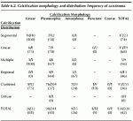
The frequency of carcinoma as a function of calcification morphology of lesions that had surgical and distribution in a study by Liberman et al biopsy is shown in Table 6.2.
Table 6.1. Mass shape and margins: frequency of carcinoma
The highest frequency of carcinoma was observed in calcifications that had linear morphology or segmental or linear distribution.
Associated Findings The mammogram report should describe associated findings, including skin or nipple retraction, skin or trabecular thickening, skin lesions, axillary adenopathy and the presence of architectural distortion.
Lesion Location Lesion location should be indicated by the breast (left, right or both), depth (anterior, middle, or posterior one-third of the breast) and location in relation to the nipple (as if you are using a clock face, facing the patient with 12:00 up, 6:00 down, 3:00 to the patient's left, and 9:00 to the patient's right: thus, 9:00 is lateral in the right breast and medial in the left breast).
Assessment Categories Every report should include an overall summary impression and give one of the BI-RADS™ assessment categories as listed in Table 6.3.
Table 6.2. Calcification morphology and distribution: frequency of carcinoma
In general, additional imaging evaluation is suggested for BI-RADS™ Category 0 lesions. Routine screening mammography is appropriate for BI-RADS™ Category 1 (normal mammogram) or 2 (benign finding). For BI-RADS™ Category 3 (probably benign) lesions, the recommendation is 6 month follow-up of the ipsilateral breast, followed by bilateral mammograms at 1 year, 2 years, and 3 years following the initial screening study. Biopsy is suggested for lesions classified as BI-RADS™ Category 4 (suspicious abnormality) and 5 (highly suggestive of malignancy).{pagebreak}
Appropriateness of Short-Term Follow-Up Mammography for "Probably Benign" Lesions The use of short-term follow-up mammography for BI-RADS™ Category 3 ("probably benign") lesions has been well supported in the literature.
{embed="ref/google-200x90"}
Examples of such lesions include single or multiple circumscribed masses and single or multiple groups of rounded or punctate calcifications.
Table 6.3. Breast imaging reporting and data system (BI-RADS™) final assessment categories
a. Assessment is incomplete Category 0 Need additional imaging evaluation: Finding for which additional imaging evaluation is needed. This is almost always used in a screening situation and should rarely be used after a full imaging workup A recommendation for additional imaging evaluation includes the use of spot compression, magnification, special mammographic views, ultrasound, etc.
The radiologist should use judgement in to how vigorously to pursue previous studies.
b.Assessment is complete - final categories Category 1 Negative: There is nothing to comment on. The breasts are symmetrical and no masses, architectural disturbances or suspicious calcifications are present.
Category 2
Benign Finding: This is also a negative mammogram, but the interpreter may wish to describe a finding. Involuting, calcified fibroadenomas, multiple secretory calcifications, fatcontaining lesions such as oil cysts, lipomas, galactoceles, and mixed-density hamartomas all have characteristic appearances and may be labeled with confidence. The interpreter might wish to describe intramammary lymph nodes, implants, etc., while still concluding that there is no mammographic evidence of malignancy.
Category 3
Probably Benign Finding - Short Interval Follow-up Suggested: A finding placed in this category should have a very high probability of being benign. It is not expected to change over the follow-up interval, but the radiologis would prefer to establish its stability. Data are becoming available that shed light on the efficacy of short interval follow-up. At the present time, most approaches ar intuitive. These will likely undergo future modification as more data accrue as to the validity of an approach, the interval required and the type of findings that should be followed.
Category 4
Suspicious Abnormality - Biopsy Should Be Considered: These are lesions that do not have the characteristic morphologies of breast cance but have a definite probability of being malignant. The radiologist has sufficient concern to urge a biopsy. If possible, the relevant probabilities should be cited so that the patient and her physician can make the decision on the ultimate course of action.
Category 5
Highly Suggestive of Malignancy - Appropriate Action Should Be Taken: These lesions have a high probability of being cancer.
In previous studies, the frequency of carcinoma for BI-RADS™ Category 3 lesions has ranged from 0.5-2.0%.
{embed="ref/stat-menu-br-cancer6"}
Sickles recommended short-term follow-up mammography in 3,184 "probably benign" lesions; cancer was subsequently discovered in 17 (0.5%).
Fifteen of the 17 cancers were diagnosed by means of interval change at follow-up mammography before they were palpable; all 17 were Stage 0 or Stage I at the time of diagnosis (one positive axillary lymph node was present in two patients). These data support the utility of the short-term follow-up mammogram in the management of "probably benign" lesions. The likelihood of cancer in this setting is extremely low, and if present it is likely that it will be diagnosed at an early and treatable stage.
More recently, Sickles has extended his work to address the question of whether patient age or lesion size should prompt immediate biopsy of nonpalpable, circumscribed, solid nodules. Of 1,403 circumscribed masses included in this study, cancer was found in 19 (positive predictive value [PPV]=1.4%). Only small differences in PPV were found for various patient age and lesion size subgroups. Even in the group of women age 50 and older, the ratio of benign to malignant was 60:1 (PPV=1.7%). These data suggest that lesion size and patient age should not deter the clinician from recommending short-interval follow-up mammography for "probably benign" lesions.
Positive Predictive Value of BI-RADS™ Categories The BI-RADS™ categories are useful predictors of malignancy. The frequency of carcinoma is 29-34% for BI-RADS™ Category 4 (suspicious abnormality) and 81-94% for BI-RADS™ Category 5 (highly suggestive of malignancy). Biopsy is generally recommended for BI-RADS™ Category 4 and Category 5 lesions. For nonpalpable masses, biopsy can be performed with needle localization and surgical biopsy or image-guided percutaneous breast biopsy.
Laura Liberman and Timothy L. Feng Breast cancer detection demonstration project: five-year summary report. CA 2003

 News
 News