Technique of Axillary Dissection
Key points
- The arm is draped free and rests on an arm board for most of the procedure.
- During dissection of the apex of the axilla, the arm can be flexed and abducted to relax the pectoralis major muscle and allow better access.
- For axillary dissection as an independent procedure, the incision is planned to maximize exposure while allowing for an excellent cosmetic result.
- While this does provide excellent exposure, scar shortening produces significant distortion, especially after breast irradiation.
The arm is draped free and rests on an arm board for most of the procedure.
During dissection of the apex of the axilla, the arm can be flexed and abducted to relax the pectoralis major muscle and allow better access. Care is taken to avoid stretching the brachial plexus.
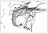
For axillary dissection as an independent procedure, the incision is planned to maximize exposure while allowing for an excellent cosmetic result. A transverse or somewhat u-shaped incision extending from the lateral margin of the pectoralis major to the anterior border of the latissimus dorsi meets these requirements (Fig. 7.9). It is important to avoid a radial incision extending from the breast to the axilla.
While this does provide excellent exposure, scar shortening produces significant distortion, especially after breast irradiation.
During modified radical mastectomy, axillary exposure is excellent after removal of the breast.
Skin flaps are elevated at the level of the axillary fascia. There are no lymph nodes superficial to this; therefore, it is unnecessary to excise the subcutaneous fat. The superior flap is elevated to the level of the transverse skin crease. A medial flap is elevated to expose the lateral margin of the pectoralis major. A lateral flap is raised to expose the anterior border of the latissimus dorsi. The extent of the inferior flap is arbitrary, as there are no bony or muscular landmarks. It is usually extended to about the fifth or sixth intercostal space (Fig. 7.9).
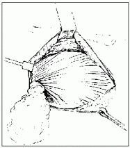
Fig. 7.7. The breast is removed along with the pectoralis fascia in a superomedial to inferolateral direction. The lateral edge of the pectoralis major is exposed. The clavipectoral fascia is then excised along the lateral margin of the pectoralis minor, from the vicinity of the axillary vein to the costal origin of the pectoralis minor. In the upper part, a neurovascular bundle containing the medial pectoral nerve and branches of the thoracoacromial vessels is found, and the nerve is preserved if possible.
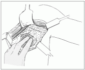
The incision is carried laterally, in front of the estimated position of the axillary vein (Fig. 7.10). At this time it is important to limit the dissection to the area below the axillary vein. Vigorous traction during dissection can easily lead the surgeon to remove a pad of fat from the front of the vein as well.
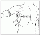
Fig. 7.8. The wound is closed over a suction drain (illustration shows a second drain in the axilla) using a subcuticular suture and steri-strips.
For a more extensive nodal clearance, the pectoralis minor muscle can be excised or divided. Unless there is extensive nodal disease, it is not usually necessary to do this. Retraction of both pectoral muscles after flexion and abduction of the arm usually allows adequate access to the apex of the axilla.
Written and revised by
Andrey Nailor, M.D.
Our doctors write and review articles for accuracy and keep them up to date.
health.am provides general information, not medical advice. Always consult a qualified health professional about your own health.
Explore health topics
More in this topic
Cancer → News
Surgical technique relieves painful spine fractures in patients with metastatic cancer
A surgical technique appears to offer quick and effective relief for debilitating spinal fractures often suffered by patients with metastatic cancer, researchers reported at the 35th Congress of the European Society for…
11 October 2010
 News
New technique in robot-assisted laparoscopic prostatectomy
Stress urinary incontinence is one of the most feared complications of radical prostatectomy. The weighted mean continence rate immediately after catheter removal following robot-assisted laparoscopic radical…
3 August 2010
 News
Studies Gauge Techniques for Measuring Breast Density -- A Predictor of Cancer
Two new studies announced this week have tested three different methods for accurately measuring breast density - the relative portion of tissue to fat in a woman's breasts and a strong indicator of breast cancer risk.…