Anal Carcinoma

Key points
- Carcinoma of the anus is a rare malignancy that usually is diagnosed at an advanced stage, in spite of being easily visible and accessible.
- An excellent review of incidence, causes, and current management of anal cancer.
- Carcinoma of the anus accounts for 1.5% of digestive system cancers [1]; when only cancers of the anorectum are considered, it represents about 5% of malignancies (range: 1% to 6%) [2, 3].
- The anal region consists of the anal canal and the anal verge (or margin).
Carcinoma of the anus is a rare malignancy that usually is diagnosed at an advanced stage, in spite of being easily visible and accessible. Its treatment has evolved from being mainly surgical to one consisting of chemotherapy (with fluorouracil and mitomycin) and radiation (megavoltage linear accelerator therapy delivering between 40 to 50 Gy). Local surgical excision is most often performed for either carcinoma in situ or microinvasive lesions of the anal margin. Radical resection is indicated for patients with residual disease following chemoradiation or for recurrent disease.
Carcinoma of the anus accounts for 1.5% of digestive system cancers [1]; when only cancers of the anorectum are considered, it represents about 5% of malignancies (range: 1% to 6%) [2, 3]. It has been estimated that 3400 patients were diagnosed with anal carcinoma in the year 2000 [1, 4]. Anal tumors tend to be most common in the sixth and seventh decades of life and are considered to have a slight female preponderance, although some series have shown a marked preponderance in females (five female patients for every male patient diagnosed) [2, 3].
The anal region consists of the anal canal and the anal verge (or margin). Considerable confusion exists in defining the anal canal. We recommend using the definition found in the 1987 edition of the International Union Against Cancer staging system [5], the 1987 edition of the Manual for Staging of Cancer of the American Joint Committee on Cancer [6], and the criteria of the World Health Organization [7]. These define the anal canal as extending from the anorectal ring to the anal verge. Using this definition, about 85% of the cancers of the anus originate in the anal canal [1, 3].
The histologic features of the anal canal and anal margin differ, reflected by the various types of anal cancer found and their management. The skin of the anal margin has a typical dermal structure except it contains an excess of sweat glands and hair. The lining of the anal canal (especially above the dentate line) contains cells that vary in morphology, including transitional cells resembling urinary tract epithelium, and a mixture of cuboidal, columnar, and squamous cells. Tumors of the anal margin can be squamous cell cancer, basal cell cancer, Bowen's disease, or Paget's disease. Cancers of the anal canal are described as squamous cell carcinoma, adenocarcinoma, cloacogenic carcinoma, basaloid carcinoma, melanoma, leiomyosarcoma, and carcinoid tumors. Cloacogenic, basaloid, and transitional tumors are considered variants of squamous cell carcinoma, which comprises more than 80% of anal canal tumors [2, 3].
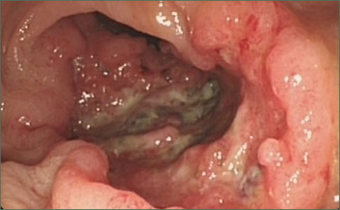
The clinical presentation of cancer of the anus is nonspecific and includes bleeding (more than 50% of patients) [3], pruritus, discharge, pain, an anal mass, and history of anal warts. A history of incontinence, change of bowel habits, pelvic pain, or rectovaginal fistula indicate an advanced lesion. Examination reveals a mass, generally ulcerated with indurated margins. The mean size of the tumor at diagnosis is about 3 cm to 4 cm . Generally, tumors of the anal canal involve only part of the circumference, whereas tumors of the margin tend to be circumferential . The diagnosis of the lesions is made by biopsy, which is best done with the patient under caudal or spinal anesthesia. The depth of the malignancy (especially for anal canal tumors) is assessed by endorectal ultrasound, which also assesses the involvement of the mesorectal lymph nodes . The site of lymph node involvement depends on the location of the tumor. Above the dentate line, lymph flow is to the pararectal and paravertebral nodes. Below the dentate line, drainage is through the inguinal and femoral nodes. The incidence of clinically positive nodes is 10% to 20% , and may be 30% to 60% for T3 or larger lesions ; 25% of lymph node metastases from T3 or larger lesions may be may be bilateral .
Written and revised by
Gerald Hurowitz, M.D.
Our doctors write and review articles for accuracy and keep them up to date.
health.am provides general information, not medical advice. Always consult a qualified health professional about your own health.
Explore health topics
More in this topic
Cancer → News
Image Analysis Project Aims to Classify Characteristics of Skin Cancer
Melanoma - the most serious form of skin cancer - varies in size, shape and severity, and although pathologists and researchers often intuitively differentiate between types of melanomas, the variations have never been…
10 September 2010
 News
Analysis Finds Sunscreens Containing Retinyl Palmitate Do Not Cause Skin Cancer
Findings refute earlier health warning issued by the Environmental Working Group Despite previous concerns about the cancer-causing potential of sunscreens containing retinyl palmitate (vitamin A), an independent…
11 August 2010
 Overview
Carcinoma of the Anus: Strategies in Management
The management of anal cancer underwent an interesting transformation over the last two decades.
21 June 2010