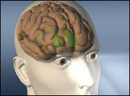
Stanford scientists see iron-containing inflammatory cells in Alzheimer's brains

Key points
- In post-mortem brain tissue from people not diagnosed with Alzheimer's, neither the iron deposits nor the scavenger cells engulfing them were present in that brain region.
- The bulk of microglia found in association with iron in the study were in an activated, inflammatory state.
- The findings also add a new suspect to the Alzheimer's disease lineup.
- "Microglia are the brain's immune cells," said Michael Zeineh, MD, PhD, assistant professor of neuroradiology and the study's lead author.
Examining post-mortem tissue from the brains of people with Alzheimer's disease, Stanford University School of Medicine investigators identified what appear to be iron-containing microglia - specialized scavenger cells that sometimes become inflammatory - in a particular part of the hippocampus, a key brain structure whose integrity is critical to memory formation.
In post-mortem brain tissue from people not diagnosed with Alzheimer's, neither the iron deposits nor the scavenger cells engulfing them were present in that brain region.
The findings, recounted in a study now available online in Neurobiology of Aging, suggest that high-field magnetic resonance imaging, in particular an advanced version called 7T MRI that uses a powerful 7-Tesla magnet, could someday be used to diagnose and monitor Alzheimer's patients earlier than is currently possible.
The findings also add a new suspect to the Alzheimer's disease lineup. A long-held hypothesis holds that the most notorious feature of Alzheimer's disease, amyloid plaques, is the main cause of the disorder. These plaques are extracellular aggregations of a small protein called beta-amyloid that are prominent in diseased patients' brains, as well as in mouse models of the disease. The other most cited key player is tau, another Alzheimer's-associated protein that abnormally aggregates into threadlike tangles inside nerve cells. Surprisingly, in the brain region of interest there was no consistent overlap between the iron-laden microglia and the amyloid plaques or tau.
"Microglia are the brain's immune cells," said Michael Zeineh, MD, PhD, assistant professor of neuroradiology and the study's lead author. In their resting state, they're like police officers in the doughnut shop, sitting down and relaxing, their guns holstered, but keeping their eyes open while placidly munching on whatever cellular debris or stray substances might come their way. If they encounter anything suspicious, though, they whirl into action. Activated microglia are like officers with their guns out and firing, Zeineh said.
Microglia inflamed
The bulk of microglia found in association with iron in the study were in an activated, inflammatory state. Alzheimer's is increasingly understood to involve brain inflammation, and groups led by Stanford researchers such as neurologists Katrin Andreasson, MD, and Tony Wyss-Coray, PhD, and neurobiologist Ben Barres, MD, PhD, have previously fingered microglia as potential suspects in the early inflammatory pathology of the disease. This study adds the new finding that inflamed, iron-associated microglia are present in the hippocampus in Alzheimer's and are observable by 7T MRI, which could advance the scientific community's understanding of the disease.
The researchers noted that this was a preliminary study performed on a small number of human brain specimens, which are generally difficult to obtain. In this case, the specimens were supplied by the study's senior author, Brian Rutt, PhD, professor of radiology.
Written and revised by
Gerald Hurowitz, M.D.
Our doctors write and review articles for accuracy and keep them up to date.
health.am provides general information, not medical advice. Always consult a qualified health professional about your own health.