Diagnostic Arguments of Ovarian Cysts in Prepubertal Girls
Key points
- Pelvic Ultrasonography Pelvic ultrasonography is central to the management of ovarian cysts.
- Plasma measurement of sex steroids (estradiol, testosterone and androgens) and basal or stimulated levels of the gonadotropins LH and FSH must be done in any endocrine syndrome.
- Tumor marker measurement is a useful diagnostic aid in lesions that are difficult to analyze by ultrasonography and in surveillance after tumor removal.
- Classically, it is carried out by a suprapubic approach with a full bladder, which allows study not only of the cyst but of all the internal genital organs.
Diagnostic Arguments of Ovarian Cysts in Prepubertal Girls
Pelvic Ultrasonography
Pelvic ultrasonography is central to the management of ovarian cysts.
Classically, it is carried out by a suprapubic approach with a full bladder, which allows study not only of the cyst but of all the internal genital organs. An analysis of the cyst itself indicates the size, shape and even volume by measurement of the three diameters, the thickness and regularity of the wall and the nature of the cyst content. An anechogenic, purely liquid image with a thin, regular wall is in favor of a functional cyst (fig. 1, 2). Volume measurement is a basis for surveillance and for the decision of possible needle aspiration.
Ultrasonography is then indispensable for topographic localization to guide aspiration. An image which is not purely liquid raises problems of differential diagnosis. If the child is symptomatic, there may be a cystic complication such as hemorrhage or ovarian torsion. The mass may be a tumor with a cystic component, raising the problem of its benign or malignant nature. Certain morphological signs are informative: volume of the tissue component, wall thickness, growths, and rigid septations with angulated junctions. Morphological color Doppler and pulsed Doppler spectral imaging study may also be useful, since these techniques provide information on the vascularization of the mass, for example, by differentiating the avascular but echogenic content of a hemorrhagic cyst from hypervascularized tumoral tissue. At a later stage, ultrasonography is used to investigate the condition of the contralateral ovary and the appearance of the uterus, its dimensions and endometrium. This information is indispensable for therapeutic management.
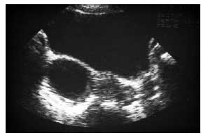
Fig. 1. Functional cyst of the right ovary: purely fluid appearance, anechogenic and homogeneous with a thin, regular wall.
In the child and adolescent, ultrasonography is limited by the impossibility of using a vaginal approach. However, suprapubic sonography is generally sufficient for appropriate diagnosis and therapeutic management of ovarian cysts. Recourse to other imaging techniques (CT, MRI) is justified essentially in two circumstances: confirmation of the multitissular nature (fat, calcification) of a dermoid cyst and assessment of the extent of a mass that is tissular rather than liquid and suspected of being malignant.
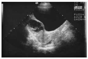
Fig. 2. Complex cyst: heterogeneous, septate appearance. The predominantly fluid component and the curved, pliable septations are in favor of a complicated benign cyst (intracystic hemorrhage in the process of resorption).
Written and revised by
David Cohen, M.D.
Our doctors write and review articles for accuracy and keep them up to date.
health.am provides general information, not medical advice. Always consult a qualified health professional about your own health.
Explore health topics
More in this topic
Women's Health → Guide
The Various Ovarian Masses
Non-Neoplastic Functional Disorders: Functional Cysts The development of simple cysts is quite common in adolescents, and the risk of malignancy is close to zero. In the pubertal period, ovaries may contain multiple…
26 September 2006
 Guide
Means of Diagnosis - Ovarian Masses in Adolescent Girls
Imaging Numerous techniques are available and play a fundamental role not only in establishing a positive diagnosis and orienting the etiology, but also in surveillance and the assessment of spread. The plain abdominal…
26 September 2006
 Guide
Management - Ovarian Masses in Adolescent Girls
In practice, the clinician is confronted by two very different situations: (1) a pelvic mass or an endocrine syndrome is discovered and complementary investigations are required for better definition of the nature of…
26 September 2006