Alzheimer Disease: From Clinical Description to a Theory of Disease and Treatment
Andrey Nailor, M.D.·Published 27 June 2011·12 min read
Key points
The story of Alzheimer disease began with a series of clinical observations that were linked to a type of brain pathology.
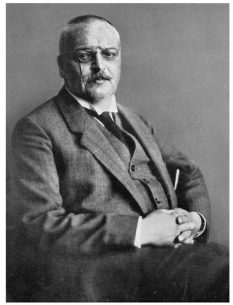
The story began on April 8, 1906, when psychiatrist Alois Alzheimer (Figure 1-1) examined the brain of Auguste D, a woman who at age 51 years had been admitted, on November 25, 1901, to the Hospital for the Mentally Ill and Epileptics in Frankfurt, Germany.
Alzheimer had seen her at the Frankfurt facility, but in 1903 had joined Emil Kraepelin at the Royal Psychiatric Clinic in Munich, where Alzheimer became head of the Anatomical Laboratory (Figure 1-2) (Goedert and Spillantini 2006).
It was here that Alzheimer received the woman's brain following her death.
The story of Alzheimer disease began with a series of clinical observations that were linked to a type of brain pathology. These clinical and pathological observations have led to theories of pathophysiology, which in turn have pointed to possible pathways to treatment. The time from disease recognition to the first symptomatic treatment was nearly 90 years. Treatments aimed at altering the disease process are still in development.
The story began on April 8, 1906, when psychiatrist Alois Alzheimer (Figure 1-1) examined the brain of Auguste D, a woman who at age 51 years had been admitted, on November 25, 1901, to the Hospital for the Mentally Ill and Epileptics in Frankfurt, Germany. Her family physician's admittance note indicated that she had been suffering for a long time from weakness of memory, persecution mania, sleeplessness, and restlessness and that she was no longer able to do any physical or mental work.
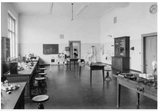
Alzheimer had seen her at the Frankfurt facility, but in 1903 had joined Emil Kraepelin at the Royal Psychiatric Clinic in Munich, where Alzheimer became head of the Anatomical Laboratory (Figure 1-2) (Goedert and Spillantini 2006).
It was here that Alzheimer received the woman's brain following her death. Grossly, he noted brain shrinkage and mild hydrocephalus ex vacuo. On applying the recently developed Bielchowsky silver stain to brain slides, he noted fibrils that stained differently from normal neurofibrils and millet seed-sized lesions containing a peculiar substance distributed throughout the outer layers of the entire cerebral cortex.
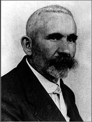
His findings were published in 1907 (Alzheimer 1907/1987); he thought the disease was a rare form of presenile dementia. By 1908, Alzheimer and his colleague Perusini had found a total of four cases of presenile dementia, all of whom had similar pathology (Maurer 2006). Only 2 years later, Kraepelin (1910) (Figure 1-3) included the eponym Alzheimer's disease in his textbook of psychiatry. Despite the fact that both plaques and tangles had been described earlier by other investigators (Drachman 2006b), the name stuck.
Belying the paucity of references in PubMed cited earlier, there was a lively discussion in the literature of the 1920s and 1930s concerning the clinical signs and symptoms of Alzheimer disease, the relationship of histopathology to clinical symptoms, and the relationship of Alzheimer disease to senile dementia (reviewed in Newton 1948).
FIGURE 1-1. Alois Alzheimer, M.D. Source. Department of Psychiatry, Ludwig Maximilian University Munich.
However, it did not become widely accepted that Alzheimer-type change in brain had a strong relationship to senile-onset dementia until the work of British pathologist J.A. N. Corsellis (1962). Corsellis performed postmortem studies of the brains of institutionalized persons with diagnoses such as schizophrenia, affective disorders, and paranoid disorders and compared them with the brains of persons having diagnoses of senile dementia or mixed senile/vascular dementia, but not with those of persons diagnosed clinically with other brain diseases. He found that 80% of the senile dementia group showed moderate to severe plaque formation.
Written and revised by
Andrey Nailor, M.D.
Our doctors write and review articles for accuracy and keep them up to date.
health.am provides general information, not medical advice. Always consult a qualified health professional about your own health.
Patients with psychiatric diagnoses had only moderate to severe plaque formation if they lived to be age 75 years or older. Neurofibrillary tangles occurred almost exclusively in persons diagnosed with senile and mixed senile/vascular dementia. These tangles tended to parallel the plaque density, and their frequency was not related to patient age. In the same year, Kay (1962) noted that a diagnosis of senile dementia was associated with markedly reduced life expectancy.
Sim and Sussman (1962) published the course of 21 cases of biopsy-proven and neuropathologically proven Alzheimer disease. They still considered the disease to be a presenile dementia, although Simchowitz, who first used the term senile plaque and had also described granulovacuolar degeneration, had written in 1914 that there was no difference between Alzheimer disease and senile dementia (Simchowitz 1914). Sim and Sussman compared the clinical picture of persons seen in years 1-4 of symptoms.
FIGURE 1-2. Alzheimer laboratory in Munich. Source. Department of Psychiatry, Ludwig Maximilian University Munich. They found amnesia and disorientation to be the earliest symptoms, dysphasia as intermediate, and apraxia and manifest dementia to occur later. They noted that in the early stages, despite poor orientation and memory loss, patients were not demented and were able to organize themselves and their environment to a remarkable degree, evidencing what we might now call mild cognitive impairment according to the criteria developed by Petersen et al. (1999).
The work of Sim and Sussman also set the stage for the later development by Rosen et al. (1984) of the Alzheimer's Disease Assessment Scale, which became one of the two standard assessment instruments in subsequent clinical trials of medication for Alzheimer disease. In 1963, electron microscopy showed that neurofibrillary tangles consisted of paired helical filaments (Kidd 1963). Three years later, Roth et al. (1966) examined the brains of patients ages 60 years and older who had been seen in a psychiatric or general hospital, had been evaluated cognitively, and did not have evidence of stroke. He came to the following three conclusions:
{pagebreak}
Far from plaques being irrelevant for the pathology of old-age mental disorder, the density of plaque formation in the brain proves to be highly correlated with quantitative measures of intellectual and personality deterioration in aged subject. The establishment of a valid measure of cerebral damage in one group of organic psychoses opens up the possibility of defining and measuring the psychological defects that show the best correlation with measures of cerebral damage. ...As the brain is an organ with a fixed cell population and such organs have long been known to be particularly liable to the changes of senescence, the facts suggest that senile plaque formation and related processes may also deserve investigation with more precise techniques than those in this study, for their possible relevance to the problems of ageing in man. (Roth et al. 1966, p. 110)
Years later, a stronger correlation was shown between cognitive impairment and reduction in synaptic density (DeKosky and Scheff 1990), but it is still not known if the age-related accumulation of amyloid plaques in the brain is a normal product of aging or a precursor of the neuritic plaques of Alzheimer disease (Knopman et al. 2003).
FIGURE 1-3. Emil Kraepelin, M.D., who named Alzheimer disease.Source. Department of Psychiatry, Ludwig Maximilian University Munich.
Roth and his group (Tomlinson et al. 1968, 1970) then compared the brains of older adults who displayed no objective evidence of cognitive impairment prior to death with those of persons with well-documented dementia.
Statistically significant between-group differences were found in cortical atrophy, ventricular dilatation, senile plaque formation, "Alzheimer's neurofibrillary change," and granulovacuolar degeneration.
FIGURE 1-4. David A. Drachman, M.D., created a pharmacologic model of age-related cognitive change.Source. Courtesy of Dr. Drachman
Because memory impairment is typically the presenting symptom of Alzheimer disease, researchers attempted to understand and treat this symptom by integrating the biology of memory with the pathophysiology of Alzheimer disease. Deutsch (1971) suggested that the cholinergic system is the biological substrate of memory. Subsequently, David Drachman (Figure 1-4) (Drachman and Leavitt 1974) showed that scopolamine blocked storage in long-term memory with relative sparing of recall and retrieval from long-term memory, a pattern similar to that of normal aging.
Searching for the relationship between neuropathology and brain function, Davies and Maloney (1976) assayed postmortem levels of enzymes associated with the neurotransmitters acetylcholine, γ -aminobutyric aid (GABA), dopamine, noradrenaline, and 5-hydroxytryptamine in 3 subjects with Alzheimer disease and 10 control subjects. Levels of choline acetyltransferase, the enzyme forming acetylcholine, and acetylcholinesterase, which hydrolyzes acetylcholine in the synapse, were dramatically reduced in the Alzheimer brains; the level of reduction coincided with the areas containing the greatest number of neurofibrillary tangles. The following year, White et al. (1977) confirmed that the presynaptic cholinergic marker choline acetyltransferase was reduced in subjects with Alzheimer disease, but also found intact postsynaptic receptor binding, suggesting that treatment with centrally acting cholinesterases might be beneficial before neuronal loss was advanced. The potential utility of this approach was underscored by the finding of massive cell loss in Alzheimer disease in basal forebrain nuclei, including the nucleus basalis of Meynert, a poorly defined group of neurons located dorsal to the optic chiasm that supplies most of the cholinergic innervation to the cerebral cortex (Whitehouse et al. 1981).
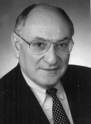
FIGURE 1-5. Kenneth L. Davis, M.D., showed memory-enhancing property of physostigmine.Source. Courtesy of Dr. Davis.
Experimentation with the anticholinesterase physostigmine by Kenneth Davis (Figure 1-5) led to the observation that it improved memory in cognitively intact individuals (Davis et al. 1978) and persons with Alzheimer disease (Davis and Mohs 1982), but the short half-life of this drug and the need for parenteral administration made it unsuitable for clinical use. At the same time, the longacting cholinesterase inhibitor tacrine was being investigated clinically, following the clinical observation that it effectively reversed anticholinergic delirium following overdose of tricyclic antidepressants (Summers et al. 1981). Following the publication of a large multicenter study by Davis et al. (1992), the U.S. Food and Drug Administration approved tacrine for the treatment of Alzheimer disease in 1993. Other cholinesterase inhibitors were later approved, and the use of tacrine gradually vanished because of the need for frequent dosing, slow titration, and hepatotoxicity. Overall, these drugs produce small, transient cognitive improvement and modest, if any, effect on disease progression, as would be expected from a symptomatic treatment.
The 1980s saw progress in elucidating the neuropathology and pathophysiology of Alzheimer disease. Glenner and Wong (1984a) sequenced from blood vessel deposits of patients with Alzheimer disease the substance that had been recognized in the core of neuritic plaques as amyloid protein (Terry et al. 1964). Based on work published the previous year showing the presence of Alzheimer disease pathology to be ubiquitous in persons with Down syndrome who had reached age 40 years, Glenner and Wong (1984b) showed that the amyloid in Down syndrome was identical to that found in patients with Alzheimer disease. Korenberg et al. (1989) later showed that the portion of chromosome that is reduplicated in trisomy 21 Down syndrome contains the amyloid precursor protein, suggesting that the cause of Alzheimer disease in these persons is overloading of the proteolytic pathway of normal amyloid precursor protein. The subsequent work in molecular genetics began with the localization of a gene for familial Alzheimer disease to chromosome 21 (St. George-Hyslop et al. 1987).
FIGURE 1-6. Robert N. Butler, M.D., first head of the National Institute on Aging. Source. Courtesy of the International Longevity Center.
{pagebreak}
It will be reviewed this sections, "Alzheimer Disease," and "The Molecular and Genetic Basis of Alzheimer Disease," as will the amyloid cascade hypothesis of Alzheimer disease, the possible neurotoxicity of amyloid beta oligomers (Walsh and Selkoe 2007), and the major risk factor for late-onset Alzheimer disease- the inheritance of the e 4 allele of the choleresterol- transporting protein apolipoprotein E (Corder et al. 1993)- and its possible relationship to the formation of neurofibrillary tangles.
Robert Katzman's (1976) editorial in the Archives of Neurology pointing out the prevalence of Alzheimer disease and its impact on life span is widely credited for sparking Alzheimer disease research in the United States (Khachaturian 2006). Congress had authorized creation of the National Institute on Aging (NIA) in 1974. By 1976, the institute was formally organized under the direction of psychiatrist Robert Butler (Figure 1-6), and in 1978, it began its program of extramural research on brain aging and Alzheimer disease under the leadership of Zaven Khachaturian (Figure 1-7). In 1979, the Alzheimer's Association (first named the Alzheimer's Disease and Related Disorders Association; ADRDA) was formed with encouragement and support from the NIA and the National Institute of Neurological and Communication Disorders and Stroke (NINCDS); Mr. Jerome Stone became President, and Drs. Robert Katzman and Carl Eisdorfer cochaired the Medical Advisory Board (Alzheimer's Association 2008).
The year 1981 saw the publication of a government industry-academia collaboration titled Strategies for the Development of an Effective Treatment for Senile Dementia (Crook and Gershon 1981) that stimulated some of the early U.S. drug trials. The title of the follow-up volume, Treatment Development Strategies for Alzheimer's Disease (Crook et al. 1986), indicated the recognition that senile dementia and Alzheimer disease were the same pathological entity. Between these two publications, on the advice of the NINCDS National Advisory Council, NINCDS and ADRDA formed the Work Group on the Diagnosis of Alzheimer's Disease to establish clinical diagnostic criteria so that the natural history of the disease could be better characterized and meaningful clinical trials undertaken. The diagnostic criteria that were formulated for possible and probable Alzheimer disease (McKhann et al. 1984) continue in use as the basis for enrollment in clinical trials.
FIGURE 1-7. Zaven Khachaturian, M.D., first associate director of the Neuroscience and Neuropsychology of Aging Program of the National Institute on Aging. Source. Courtesy of Dr. Khachaturian.
Morris and Rubin (1991) reported that these criteria have 80%-90% accuracy. A recent review of autopsy cases (Ranginwala et al. 2007) finds this accuracy rate essentially unchanged despite advances in immunohistochemistry that now enable more ready detection of Lewy bodies and tauopathies other than the neurofibrillary tangles of Alzheimer disease.
In 1986, a grant from NIA enabled the creation of the Consortium to Establish a Registry for Alzheimer's Disease (CERAD) to standardize the clinical, neuropsychological, and neuropathological evaluation of persons with suspected Alzheimer disease. In this effort, led by Dr. Albert Heyman of Duke University, patients with Alzheimer disease and control subjects without dementia were recruited from 24 NIA-sponsored Alzheimer's disease centers and from other university programs in the United States. Subjects were examined at entry and annually thereafter, to observe the natural progression of Alzheimer disease. Autopsy examination of the brain was included, to the extent possible. The major standardized instruments developed by CERAD are now used by many Alzheimer disease research centers in the United States and abroad, by physicians in clinical practice, and in population-based surveys. Data were obtained on more than 1,000 nationally distributed white and black patients with Alzheimer disease and nearly 500 control subjects without dementia. In this series, the clinical diagnosis of Alzheimer disease based on NINCDS-ADRDA criteria was confirmed in 87% of autopsied cases. The CERAD database is still available to investigators.
In 1991, the Alzheimer's Disease Cooperative Study was funded by NIA under the direction of the late Leon Thal (Figure 1-8) to provide an infrastructure for clinical trials of substances that have included estrogen and nonsteroidal anti-inflammatory agents.
Most recently (2004), the Alzheimer's Disease Neuroimaging Initiative was established with resources from the National Institute on Aging, the National Institute for Biomedical Imaging and Bioengineering, the Foundation for the National Institutes of Health, the Alzheimer's Drug Discovery Foundation, and the Alzheimer's Association to examine how brain imaging technology can be used with other biological marker tests to measure the progression of mild cognitive impairment and early Alzheimer disease. This information will then be used to aid future clinical trials by providing a standard assessment tool to measure treatment effects (Mueller et al. 2005).
###
Myron F. Weiner, M.D. Clinical Professor of Psychiatry and Neurology, Aradine S. Ard Chair in Brain Science, Dorothy L. and John P. Harbin Chair in Alzheimer's Disease Research, University of Texas Southwestern Medical Center at Dallas, Texas ###
REFERENCES
Adams RD, Victor M: Principles of Neurology, 4th Edition. New York, McGraw-Hill, 1989
Alzheimer A: About a peculiar disease of the cerebral cortex (1907). Translated by Jarvik L, Greenson H. Alzheimer Dis Assoc Disord 1:7-8, 1987
Alzheimer's Association: Milestones. 2008. Available at: http://www.alz.org/about_us_milestones.asp. Accessed April 21, 2008.
American Psychiatric Association: Diagnostic and Statistical Manual: Mental Disorders. Washington, DC, American Psychiatric Association, 1952
American Psychiatric Association: Diagnostic and Statistical Manual of Mental Disorders, 2nd Edition. Washington, DC, American Psychiatric Association, 1968
American Psychiatric Association: Diagnostic and Statistical Manual of Mental Disorders, 3rd Edition, Revised. Washington, DC, American Psychiatric Association, 1980
American Psychiatric Association: Diagnostic and Statistical Manual of Mental Disorders, 4th Edition. Washington, DC, American Psychiatric Association, 1994
American Psychiatric Association: Diagnostic and Statistical Manual of Mental Disorders, 4th Edition, Text Revision. Washington, DC, American Psychiatric Association, 2000
Aretaeus: The Extant Works of Aretaeus, the Cappadocian. Edited by London F. London, Syndenham Society, 1861
Bachman DL, Wolf PA, Linn, R, et al: Prevalence of dementia and probable senile dementia of the Alzheimer type in the Framingham Study. Neurology 42:115-119, 1992
Bleuler E: Textbook of Psychiatry. Translated by Brill AA. New York, Macmillan, 1924
Boeve BF: A review of the non-Alzheimer dementias. J Clin Psychiatry 67:1985-2001, 2006Dementia and Alzheimer Disease 15
Butler RN: Age-ism: another form of bigotry. Gerontologist 9:243-246, 1969

 News
 News
 News