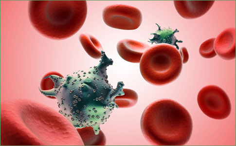
Scientists image a single HIV particle being born

Key points
- A mapmaker and a mathematician may seem like an unlikely duo, but together they worked out a way to measure longitude – and kept millions of sailors from getting lost at sea.
- This work, published in the May 25 advanced online issue of Nature, may not only prove useful in developing treatments for the millions around the globe still living with the lethal virus but the technique created to image its assembly may also change the way scientists think about and approach their own research.
- “The use of this technique is almost unlimited,” says Nolwenn Jouvenet, a postdoc who spearheaded this project under the direction of HIV expert Paul Bieniasz and cellular biophysicist Sandy Simon, who has been developing the imaging technique since 1992.
- Unlike a classical microscope, which shines light through a whole cell, the technique called total internal reflection microscopy only illuminates the cell’s surface where HIV assembles.
A mapmaker and a mathematician may seem like an unlikely duo, but together they worked out a way to measure longitude – and kept millions of sailors from getting lost at sea. Now, another unlikely duo, a virologist and a biophysicist at Rockefeller University, is making history of their own. By using a specialized microscope that only illuminates the cell’s surface, they have become the first to see, in real time and in plain view, hundreds of thousands of molecules coming together in a living cell to form a single particle of the virus that has, in less than 25 years, claimed more than 25 million lives: HIV.
This work, published in the May 25 advanced online issue of Nature, may not only prove useful in developing treatments for the millions around the globe still living with the lethal virus but the technique created to image its assembly may also change the way scientists think about and approach their own research.
“The use of this technique is almost unlimited,” says Nolwenn Jouvenet, a postdoc who spearheaded this project under the direction of HIV expert Paul Bieniasz and cellular biophysicist Sandy Simon, who has been developing the imaging technique since 1992. “Now that we can actually see a virus being born, it gives us the opportunity to answer previously unanswered questions, not only in virology but in biology in general.”
Unlike a classical microscope, which shines light through a whole cell, the technique called total internal reflection microscopy only illuminates the cell’s surface where HIV assembles. “The result is that you can see, in exquisite detail, only events at the cell surface. You never even illuminate anything inside of the cell so you can focus on what you are interested in seeing the moment it is happening,” says Simon, professor and head of the Laboratory of Cellular Biophysics.
When a beam of light passes through a piece of glass to a cell’s surface, the energy from the light propagates upward, illuminating the entire cell. But when that beam is brought to a steeper angle, the light’s energy reflects off the cell’s surface, illuminating only the events going on at its most outer membrane. By zeroing in at the cell’s surface, the team became the first to document the time it takes for each HIV particle, or virion, to assemble: five to six minutes. “At first, we had no idea whether it would take milliseconds or hours,” says Jouvenet. “We just didn’t know.”
“This is the first time anyone has seen a virus particle being born,” says Bieniasz, who is an associate professor and head of the Laboratory of Retrovirology at Rockefeller and a scientist at the Aaron Diamond AIDS Research Center. “Not just HIV,” he clarifies, “any virus.”
To prove that what they were watching was virus particles assembling at the surface (rather than an already assembled virion coming into their field of view from inside the cell), the group tagged a major viral protein, called the Gag protein, with molecules that fluoresce, but whose color would change as they packed closer together. Although many different components gather to form a single virion, the Gag protein is the only one necessary for assembly. It attaches to the inner face of the cell’s outer membrane and when enough Gag molecules flood an area, they coalesce in a way that spontaneously forms a sphere.
Written and revised by
David Cohen, M.D.
Our doctors write and review articles for accuracy and keep them up to date.
health.am provides general information, not medical advice. Always consult a qualified health professional about your own health.
Explore health topics
More in this topic
Infections, HIV & STIs → News
Scripps Research scientists identify key interaction in hepatitis C virus
Scientists from the Florida campus of The Scripps Research Institute have identified a molecular interaction between a structural hepatitis C virus protein (HCV) and a protein critical to viral replication. This new…
29 December 2010
NewsGladstone scientists link hepatitis C virus infection to fat enzyme in liver cells
Scientists at the Gladstone Institute of Virology and Immunology (GIVI) have found that an enzyme associated with the storage of fat in the liver is required for the infectious activity of the hepatitis C virus (HCV).…
12 October 2010
NewsNIH scientists find more health benefits from starting HIV treatment early
HIV-infected individuals who begin antiretroviral therapy (ART) soon after acquiring the virus may have stronger immune responses to other pathogens than HIV-infected individuals who begin ART later, a new study from…