
Fig. 2. Sonogram by abdominal route depicting the polycystic appearance of the ovaries in an adolescent girl with PCOS. Note that both ovarian sections are greater than the uterine section. In this representative case, the increased number of subcapsular follicles and the stromal hyperplasia are easily seen.
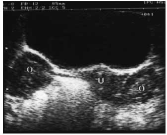
Fig. 2. Sonogram by abdominal route depicting the polycystic appearance of the ovaries in an adolescent girl with PCOS. Note that both ovarian sections are greater than the uterine section. In this representative case, the increased number of subcapsular follicles and the stromal hyperplasia are easily seen.