
Figure 42-11
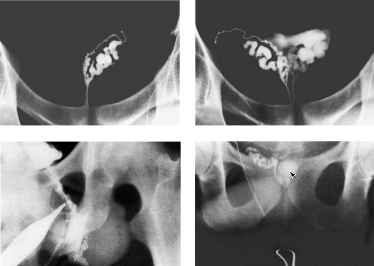
Figure 42-11. Vasography demonstrating (upper left) normal vas and seminal vesicles and (upper right) normal reflux into the bladder, confirming duct patency. Epididymal imaging showing extravasation (lower left) that demonstrates the difficulty of vasal dye injection toward the testis. Lower right:
Azoospermia from a midline ejaculatory duct cyst is shown with contrast
material filling the cyst (arrow).