
Figure 6-1 - Nuclear imaging during stress testing.
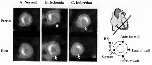
Nuclear imaging during stress testing. (a) Normal scan. Both rest and stress images demonstrate
homogeneous perfusion of the myocardium. (b) Ischemia. The stress image demonstrates a large inferior wall
perfusion defect that "fills in" in the rest images. (c) Infarction. Both rest and stress images demonstrate a large
inferior wall perfusion defect.