
Figure 20-4. MRI scan of invasive bladder carcinoma:
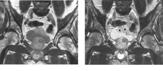
Figure 20-4. MRI scan of invasive bladder carcinoma: Left: T1-weighted image; Right: T2-weighted image. Bladder wall invasion is best assessed on T2-weighted images because of heightened contrast between tumor (asterisks) and detrusor muscle along with ability to detect interruption of the thin high-intensity line representing normal bladder wall. The heterogeneous appearance of the prostate (arrow) on the T2-weighted image owes to benign prostatic hypertrophy, confirmed at cystectomy.