Figure 18-6
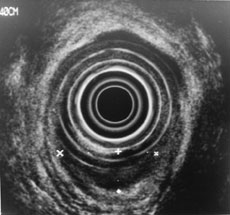
Figure 18-6. A T1 lesion is depicted. Note the hypoechoic focal mass that involves the mucosal and submucosal layers only.
 Health.am
Figure 18-6. A T1 lesion is depicted. Note the hypoechoic focal mass that involves the mucosal and submucosal layers only.