
Figure 18-2
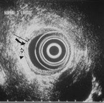
Figure 18-2. Endoscopic ultrasound image of the normal five-layered esophagus. Two small arrows: The inner-most layer corresponds to an interface echo and the mucosa. Single long arrow: The second layer corresponds to the mucosa (including the muscularis mucosa). Curved solid arrow: The third layer corresponds to the submucosa. Open arrow: The fourth layer corresponds to the muscularis propria. Solid arrowhead: The fifth outer-most layer corresponds to an interface echo and the adventitia (serosa equivalent). The five-layered appearance is essentially the same throughout the gastrointestinal tract. Because the esophagus lacks a serosa, the fifth echogenic layer in the esophagus represents the interface echo with the adventitia.