Gerald Hurowitz, M.D.·Published 29 April 2008·21 min read
Key points
Fecal incontinence (FI), the involuntary loss of formed stool, can be a social as well as a hygiene problem and is often devastating for patients and their caretakers.
FI is the involuntary leakage of stool or gas from the rectum.
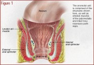
Continence is maintained by the integrated action of the structural and functional portions of the anorectal unit, which is comprised of the internal anal sphincter (IAS), external anal sphincter (EAS), striated muscles of the puborectalis (PBR), and intact neurosensory pathways (see Figure 1).
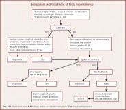
Patients with predisposing medical conditions should trigger a specific line of questions by the practitioner regarding FI (see the algorithm).
Fecal incontinence (FI), the involuntary loss of formed stool, can be a social as well as a hygiene problem and is often devastating for patients and their caretakers. Current data, which are probably underestimated, indicate that the occurrence is remarkably high. The etiology of FI is multifactorial; risk factors include advancing age, previous obstetric trauma, diabetes, fecal impaction, stroke, and dementia. The management of FI in the elderly depends on etiologic factors. However, there are many treatment options for sufferers of FI including bulking agents, antidiarrheals, anticholinergics, biofeedback, surgery for sphincter defects, and sphincter bulking devices. The appropriate treatment can be guided by a thorough workup of these patients and can result in a significant improvement in quality of life.
Fecal incontinence is an underreported problem in the general population; it is especially common in elderly persons (aged ?65 years) residing in the community or in long-term care settings. It affects more women than men during younger years, but this differential narrows with age. Physiological changes such as sphincter muscle and sensory abnormalities in the anorectal region contribute to this problem, as do factors such as dementia, physical disability, and fecal impaction.
Treatment with biofeedback is feasible in many elderly patients. Those with advanced dementia or physical disability may benefit from a bowel habit training program. Selected patients may require surgical sphincter repair. Minimally invasive techniques such as radiofrequency energy application offer promising future treatment options. The purpose of this review is to provide current information on fecal incontinence and its management.
Roach M, Christie JA. Fecal incontinence in the elderly. Geriatrics. 2008;63(2):13-22.
Fecal incontinence (FI) is a socially stigmatizing disease that disproportionately affects adults aged 65 and older. Its true prevalence remains largely unknown because of underreporting, but its predominance in the elderly population is undisputed. Nursing home residents bear the burden of this disease. Furthermore, studies have shown an excess in morbidity among elderly persons with FI living at home. The most effective way to address FI is to identify and treat the etiologic factors. This review focuses on the pathophysiology, diagnosis, and treatment of FI in elderly individuals.
Prevalence
Written and revised by
Gerald Hurowitz, M.D.
Our doctors write and review articles for accuracy and keep them up to date.
health.am provides general information, not medical advice. Always consult a qualified health professional about your own health.
FI is the involuntary leakage of stool or gas from the rectum. Clinical manifestations may range from fecal soiling to complete loss of stool from the rectum. The personal impact of FI is profound and far-reaching because of the social isolation that ensues as an individual's level of functioning in daily life is governed by proximity to a toilet. This disease frequently results in the institutionalization of the elderly because of the difficulty families experience in coping with the disease. It is a marker of poor health and increased mortality. FI is associated with an increase in depression and anxiety among patients that inevitably results in a true or perceived degradation in quality of life.
The exact incidence and prevalence of FI is unknown because of the reluctance of patients to discuss this disorder and the failure of physicians to inquire about it. Nonetheless, its prevalence varies with the study population, 2% to 17% in community-dwelling elderly, 50% to 65% in nursing home elderly, and 33% in hospitalized elderly.
Double incontinence (FI and urinary incontinence [UI]) has been found to be more common than either entity alone, with 50% to 70% of patients with UI also carrying the diagnosis of FI. This striking concordance of UI and FI suggests a similar underlying physiologic mechanism, which may include damage to the pelvic floor as a result of trauma or surgery, neurologic disorders, functional impairment, or dementia. Multiple studies have shown that dementia and immobility play a contributory role in UI and FI.
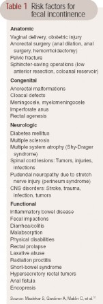
Table 1 Risk factors for fecal incontinence Higher prevalence rates are seen among patients with predisposing conditions such as diabetes mellitus (20%), irritable bowel syndrome (20%), stroke (new onset, 30%; 15% at 3 years poststroke), multiple sclerosis (51%), traumatic spinal cord injury (27%), and spina bifida (80%). These conditions are known to affect mobility as well as cause diarrhea or fecal impaction, all of which are risk factors for incontinence (see Table 1). The higher prevalence among the elderly and nursing home residents may be attributable to clinical and functional factors associated with age such as a higher incidence of constipation, dependence and immobility, cognitive impairment, polypharmacy, and general poor health.
Physiology of continence
Continence is maintained by the integrated action of the structural and functional portions of the anorectal unit, which is comprised of the internal anal sphincter (IAS), external anal sphincter (EAS), striated muscles of the puborectalis (PBR), and intact neurosensory pathways (see Figure 1). Continence is influenced by the consistency and volume of stool delivered to the rectum, the capacity and compliance of the rectum, anorectum sensation, and resting tone. Failure of any component of the anorectal unit may lead to FI when other components/mechanisms are unable to compensate for the defect in the anorectal unit.
The IAS is a 0.3- to 0.5-cm distal continuation of the circular smooth-muscle layer of the rectum. It maintains approximately 70% to 80% of anal resting tone. With increasing age, resting pressure tends to decrease because of gradual degeneration of the IAS. Studies have shown that there is a 30% to 40% decrease in sphincter pressures in adults older than 70 when compared to adults younger than 30. Whether this is attributable to medication or age-related changes in pelvic floor dynamics is unclear. There is evidence that in all age-groups, lower squeeze pressure affects women more than men, with an accelerated diminution after menopause. This may be related to the estrogen-deficient state of menopause, because estrogen receptors have previously been identified in the human stroma and smooth muscles of the anal canal. However, a recent review of the literature failed to show statistically significant gender differences, citing only age-related variation in squeeze pressure.
The EAS is a 0.6- to 1.0-cm expansion of the levator ani muscles (see Figure 1). It circles the IAS and is a tonically active striate muscle that contributes to the remaining 20% to 30% of resting tone. The IAS maintains anal continence at rest while the EAS, PBR, and levator ani assist by voluntary contraction when necessary to maintain continence. The PBR is the proximal continuation of the EAS and it passes posteriorly around the lower part of the rectum to form a sling that creates the anorectal angle. The EAS and PBR are innervated by the distal fibers of the pudendal nerve with some contribution from S4 to the PBR. Progressive neuropathic changes to the external anal sphincter may worsen with aging as evidenced by an increase in length in the motor unit potentials responsible for sphincter contraction. Anal closure is also maintained by the anal mucosal folds and the vascular cushions that expand to keep a tight seal and may contribute 10% to 20% of anal resting pressure (see Figure 1).
Figure 1 The anorectal unit is comprised of the structures shown here, as well as striated muscles of the puborectalis and intact neurosensory pathways. (Illustration: Kevin Somerville)
{pagebreak}Pathophysiology of FI - Fecal incontinence in the elderly
FI in the elderly is often multifactorial. It may be the result of structural and functional decompensation. Impairment of the anorectal unit, such as weakness from prolonged straining secondary to constipation or overt anal tears seen after vaginal delivery in women (35%), are common causes of FI. The injury sustained from obstetric trauma is often delayed in onset; many women do not manifest symptoms until their 50s.
Idiopathic FI is an entity frequently ascribed to women with idiopathic denervation of the PBR muscle and the EAS. Many of these women are multiparous and lack other predisposing factors. Other factors implicated in the cause of idiopathic FI are nerve entrapment, descending perineum syndrome, as well as central and peripheral neuropathy.
The consistency and volume of stool may be a contributing factor in the development of FI. Diarrhea and constipation exacerbate FI. Chronic diarrhea from illnesses such as inflammatory bowel disease, irritable bowel syndrome, bacterial overgrowth, short-bowel syndrome, celiac disease, bile acid malabsorption, and diabetic diarrhea overwhelms the anorectal unit and places individuals at risk for FI.
Fecal impaction is reported to occur in more than 40% of elderly patients admitted to the hospital. Read et al found no significant difference in anal pressures and maximum basal and squeeze sphincter pressures between impacted individuals and controls. However, perianal sensation was found to be absent or impaired in 74% and 77% of impacted patients, respectively, with only 29% of patients being able to distinguish between gas and liquid in the anal canal. It is believed that impaired sensation prevents patients from guarding against the relaxation of the EAS that occurs with rectal distention, preventing the voluntary contraction of the external sphincter and PBR muscle unit. With the administration of laxatives, FI is further aggravated by the leakage of fecal material around the impacted mass, leading to paradoxical diarrhea.
Clinical evaluation
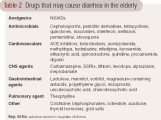
Patients with predisposing medical conditions should trigger a specific line of questions by the practitioner regarding FI (see the algorithm). Obtaining information on the frequency and nature of FI may help clinical practitioners understand the severity of incontinence and its impact on the patient's quality of life. History taking should include onset, frequency, characterization of bowel habits, consistency of stool, awareness of the desire to defecate, and risk factors. Travel history should also be discussed as well as medication history including recent antibiotic use (see Table 2). Patients may report FI in the setting of loose, copious stool that may result from the inability of the rectum to hold large volumes of loose stool.
Table 2 - Evaluation and treatment of fecal incontinence
A multisystem physical examination should include a complete anorectal examination conducted in both the left lateral decubitus and seated positions. Digital rectal exam (DRE) allows for a subjective assessment of resting and squeeze pressures (the pressure generated by the patient when asked to contract the sphincter for ≥ 30 seconds), presence of a mass, fecal impaction, scars, a thin perineal body, anal fissures, and pain on examination. The positive predictive value of the DRE for identifying resting and squeeze pressures is 67% and 81%, respectively.
The normal anterior and upward lift of the PBR sling, perianal sensation, and elucidation of the anal wink provides good assessment of neuromuscular functioning. The anal wink is tested by stroking the skin lateral to the anal opening, which should elicit contraction of the anus in normal patients. The absence of this reflex suggests neural damage (pudendal nerves and S4 nerve roots). The discovery of fecal impaction may predispose to overflow incontinence in the elderly and points to the need for disimpaction with an enema and institution of a good bowel regimen. Scars from prior surgery and fistulas may be evident as well as hemorrhoids, and ballooning of the perineum, which may be a sign of a weak pelvic floor. Further, during the performance of the Valsalva's maneuver, a gaping or open anus suggests rectal prolapse or a neurologic disorder.
Diagnostic testing
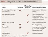
The use of diagnostic testing in the elderly should be governed by the patient's comorbidities, expected life span, symptom severity, suspected etiology, and response to medical management. The primary diagnostic modalities available for the evaluation of FI include flexible sigmoidoscopy, colonoscopy, anorectal manometry, endoanal ultrasound (EAUS), evacuation proctography (defecography), pelvic MRI, and electromyography of the external sphincter (see Table 3). These procedures are performed in specialized clinical laboratories under the auspices of a gastroenterologist, radiologist, colorectal surgeon, or neurologist. To ensure quality control and patient safety, the American Neurogastroenterology and Motility Society is currently considering the formal registration and listing of all laboratories that regularly perform these clinical tests.
Flexible sigmoidoscopy and colonoscopy Flexible sigmoidoscopy or colonoscopy is recommended in most elderly patients with FI. Visualization of the rectosigmoid mucosa should be considered to rule out structural problems such as internal hemorrhoids, fistulas, fissures, neoplasm, or inflammation associated with ischemia, colitis, and pseudomembranes. Sigmoidoscopy can be done at the bedside in patients with limited mobility or varying degrees of dementia. Colonoscopy should be performed in patients without significant morbidities, as this procedure is invasive and requires sedation.
Anorectal manometry This allows for the assessment of IAS resting pressure, maximum squeeze pressure, the sphincter length, and the integrity of the rectoanal inhibitory reflex and rectal sensation. Measurements are taken as a rectal pressure catheter with a balloon at the tip is slowly withdrawn in 0.5- to 1-cm intervals from the rectum to the anal verge. Measurements are also taken during various simulated events such as the Valsalva's maneuver and postponement of defecation. Anal resting and squeeze pressures are frequently observed to be reduced in FI. Normal values are influenced by age, gender, and the technique utilized, and they need to be compared against normal values obtained in age- and gender-matched subjects. The anal canal length and maximal squeeze pressures are greater in men when compared to parous women. However, this difference is not seen in the resting anal pressure of nulliparous women. Studies have shown that maximum squeeze pressures have good sensitivity and specificity for differentiating between FI and controls. Interestingly, in most cases, there is little improvement in manometric measures with successful treatment of FI. Anal manometry requires patients to be able to follow directions for accurate completion of the test, so demented patients are not good candidates.
Rectoanal sensory testing The rectal balloon sensory testing allows assessment of the awareness of rectal distention. During rectal balloon distention, thresholds for first perception, desire to defecate, and maximal tolerable volume are measured. In normal controls, the reflex is seen after instillation of 10 to 30 mL of air and induces a desire to pass flatus after 40 to 50 mL of air is instilled. Rectal compliance as measured by balloon distention assesses the patient's ability to postpone defecation to a convenient time. Patients must be able to follow commands for the completion of this test.
Table 3 Diagnostic studies for fecal incontinence
Endoanal ultrasound EAUS is useful for assessing morphologic anomalies of the anal sphincters such as sphincter thinning, scars, defects, and fistulae. Three-dimensional images are obtained using a 7-MHz or 10-MHz transducer providing a 360-degree view of the anal canal. It allows accurate visualization of the IAS; however, assessment and interpretation of EAS may be more operator dependent and confounded by normal anatomic variants as well as interobserver variability. In experienced hands, the sensitivity and specificity of this method approaches 100% in identifying defects of both the EAS and IAS. The patient's mental status does not seem to affect the result of this clinical test.
Proctography During dynamic proctography, anorectal anatomy and pelvic floor motion are recorded at rest, coughing, squeezing, and straining to expel barium from the rectum. Additionally, the anorectal angle and position of the anorectal junction are tracked during these maneuvers as well as retention and evacuation of contrast material. This test may be used to assess the presence of excessive perineal descent, a rectocele or enterocele, or internal rectal intussusceptions. This technique is rapidly being replaced by dynamic MRI because the technique for conducting MRI and interpretation of data are more standardized. Patients must be able to follow the commands for completion of this test.
Pelvic MRI This modality allows visualization of the anal sphincter anatomy and global pelvic floor motion in real time. Dynamic imaging of the pelvic floor is done as a patient squeezes the sphincter muscles to evacuate ultrasound gel from the rectum or perform the Valsalva's maneuver. Examination may be performed in a supine or seated position. The latter technique is superior to EAUS for detecting rectal intussusceptions. Endoanal MRI has been shown to be equal or superior to EAUS for assessing the EAS. It also allows detection of PBR atrophy in FI. Added benefits include dynamic assessment of global pelvic floor motion and visualization of the bladder and genital organs.
Needle electromyography of the external sphincter The EAS and PBR muscles are assessed by needle insertions and measurement of insertional activity at rest, motor unit potential amplitude and duration, percentage of polyphasia, and recruitment following mild to moderate voluntary contraction. EAUS, however, has largely replaced it for identifying sphincter defects.
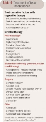
{pagebreak}Treatment options - Fecal incontinence in the elderly Diarrhea and constipation are independent risk factors for fecal incontinence. Therefore, medical management of FI is directed toward identifying and treating the underlying disorders causing diarrhea or constipation (see Table 4). Surgical and behavioral treatments address the integrity and strength of the pelvic floor muscles as well as sensory dysfunction of the rectum associated with neurologic and anatomic defects. When both treatment modalities fail to restore bowel control or improve quality of life, therapy is targeted toward providing symptom relief.
Medical management Supportive treatment should begin with simple lifestyle modifications such as avoiding certain foods and bowel habit training. Treatment of underlying causes of diarrhea or constipation may result in cure in a small subset of patients. Avoiding caffeine, a gastrocolonic stimulant, after meals as well as brisk physical activity immediately postprandially may lessen urgency and diarrhea. In all patients, especially the institutionalized, good skin hygiene is important to prevent skin breakdown. Frequent cleansing, changing of undergarments, and use of barrier creams such as silver sulfadiazine, zinc oxide, and calamine lotion may prevent skin excoriation. Scheduled or ritualized toileting, instituting cognitive training, and providing easily accessible facilities such as bedside commodes and bedpans may prove helpful in decreasing frequency of soiling and accidents.
Table 4 Treatment of fecal incontinence
Bulking agents such as soluble fiber may be used to increase stool bulk, but there is little evidence to support this approach. Antidiarrheal agents such as loperamide are the first line of pharmacologic agents employed in persistent diarrhea. Loperamide, a synthetic opioid, decreases intestinal motility and secretion. A placebo-controlled trial of loperamide showed reduced frequency of incontinence, improved stool urgency, and increased colonic transit time as well as an increase in anal resting sphincter pressure and reduced stool weight. Loperamide taken before social occasions may help incontinent patients avoid accidents and thus encourage participation in social activities; however, common side effects include constipation and dependency.
Diphenoxylate/atropine is an alternative drug that is used with variable results. Codeine phosphate does provide benefit but at the potential cost of increased drowsiness and addiction. Cholestyramine/colestipol may be used in patients with ileal resection for whom bile salt malabsorption may be the underlying disorder. Other agents such as estrogen and sodium valproate have been tried with mixed results. Agents such as tricyclic antidepressants have been used empirically to improve symptoms for patients with idiopathic FI. In an open-label study of low-dose amitriptyline in the treatment of patients with idiopathic FI (mean age, 66 years), 89% of patients reported improvement in symptoms and reduced number of bowel movements per day (P*gt; .001). Phenylephrine gel has been shown to increase resting anal canal pressure and FI scores for patients with ileoanal reservoir pouch and FI.
In patients with constipation, fecal impaction, and overflow incontinence, regular colonic evacuation with enemas, digital stimulation with bisacodyl or glycerin suppositories, fiber supplementation, and selective use of oral laxatives may prove helpful.
Biofeedback Biofeedback is usually the first line of treatment if medical management fails. It is based on the principles of operant conditioning, and is used in cooperative patients with weak sphincters and/or impaired rectal sensation. Using a rectal balloon anal manometry device, patients are taught to contract the EAS when they perceive balloon distention. The goal is to improve contraction of the EAS in response to rectal distention, and visual, auditory, or verbal feedback techniques may be employed. Most patients seem to require 4 to 6 training sessions for a sustained response. Most studies revealed short-term subjective improvements of symptoms, but without improvement in objective anorectal measurements. Some studies do report improvement in voluntary squeeze pressure, rectoanal coordination, and rectal sensation and capacity. The evidence suggests that biofeedback has been found in the short term to improve symptoms and restore quality of life for patients aged 50 and older.
The presence of severe FI, pudendal neuropathy, and neurologic disorders portends a poor prognosis when treating with biofeedback. No reported randomized, controlled trials have compared biofeedback to other modalities including surgery. The technique remains incompletely standardized, and defining clinical improvement remains a challenge. However, given its safety profile and ease of application, biofeedback should be offered to all patients with intact cognitive function in whom supportive medical therapies have failed.
Anal plug devices, injectable sphincter-bulking agents (eg, autologous fat, collagen and implantable microballoons, carbon-coated beads and silicone), and electrical stimulation of the sacral nerve should be considered experimental therapies. Disposable anal plugs for temporary occlusion of the anal canal may be useful for patients with impaired anal canal sensation and neurologic disease and for institutionalized or immobilized patients. Short-term improvement has been shown with bulking of the anal sphincter to augment surface area and provide a tighter seal for the anal canal. The use of sacral nerve stimulation as an alternative treatment warrants further study.
{pagebreak}Surgery - Fecal incontinence in the elderly Surgical therapy is usually indicated for anatomic defects in the pelvic floor musculature and anal sphincters. Sphincteroplasty with overlapping anterior sphincter repair is the primary surgical procedure in patients with EAS defects commonly seen as a result of obstetric trauma. If an IAS defect is identified, then IAS imbrication (ie, overlapping of tissue layers) is performed. During dynamic graciloplasty, the fast twitch fatigable skeletal muscle is transformed into a slow twitch fatigable muscle that can provide sustained sphincterlike muscle response by an implanted pacemaker. In a 2002 systematic review of patients who had undergone dynamic graciloplasty, clinical improvements ranged between 38% and 90%. An alternative to stimulated gracilis muscle transposition is the artificial bowel sphincter (ABS), which like dynamic graciloplasty is placed around the native sphincter via perineal tunnels. Revision of both procedures approaches 50%. Currently, there is insufficient evidence on safety and effectiveness of ABS for FI, and it should be considered experimental. Experience with antegrade continence enema through a cecostomy button or appendicostomy is limited, but short-term results show a benefit of 40% to 60% in some series.
Summary
The management of FI in the elderly should employ an individualized and minimally invasive approach that is both practical and convenient. Attention to good nursing care with the application of a skin protectant to prevent ulcers, institution of motivational techniques, and assistance in keeping the elderly mobile is very important. Ritualized bowel evacuation/emptying regimens will prevent fecal impaction and overflow diarrhea. Fortunately, there are several medical as well surgical options for patients with FI. The ultimate goal is to improve quality of life and reduce morbidity and mortality in patients suffering with this potentially psychosocially and physically debilitating disorder.
References
1. Nelson R, Furner S, Jesudason V. Fecal incontinence in Wisconsin nursing homes: prevalence and associations. Dis Colon Rectum. 1998;41(10):1226-229.
2. Nelson R, Norton N, Cautley E, et al. Community-based prevalence of anal incontinence. JAMA. 1995;274(7):559-561.
3. Edwards NI, Jones D. The prevalence of faecal incontinence in older people living at home. Age Ageing. 2001;30(6):503-507.
4. Ouslander JG, Kane RL, Abrass IB. Urinary incontinence in elderly nursing home patients. JAMA. 1982;248(10):1194-1198.
5. Kok AL, Voorhorst FJ, Burger CW, et al. Urinary and faecal incontinence in community-residing elderly women. Age Ageing. 1992;21(3):211-215.
6. Bliss DZ, Jung HJ, Savik K, et al. Supplementation with dietary fiber improves fecal incontinence. Nurs Res. 2001;50(4):203-213.
7. Ouslander JG, Palmer MH, Rovner BW, et al. Urinary incontinence in nursing homes: Incidence, remission and associated factors. J Am Geriatr Soc. 1993;41(10):1083-1089.
8. Harari D, Coshall C, Rudd AG, et al. New-onset fecal incontinence after stroke: prevalence, natural history, risk factors, and impact. Stroke. 2003;34(1):144-150.
9. Hinds JP, Eidelman BH, Wald A. Prevalence of bowel dysfunction in multiple sclerosis. A population survey. Gastroenterology. 1990;98(6):1538-1542.
10. Krogh K, Nielsen J, Djurhuus JC, et al. Colorectal function in patients with spinal cord lesions. Dis Colon Rectum. 1997;40(10):1233-1239.
11. Okamoto GA, Lamers JV, Shurtleff DB. Skin breakdown in patients with myelomeningocele. Arch Phys Med Rehabil. 1983;64(1):20-23.
12. Talley NJ, O'Keefe EA, Zinsmeister AR, et al. Prevalence of gastrointestinal symptoms in the elderly: A population-based study. Gastroenterology. 1992 ;102(3):895-901.
13. Drossman DA, Sandler RS, Broom CM, et al. Urgency and fecal soiling in people with bowel dysfunction. Dig Dis Sci. 1986;31(11):1221-1225.
14. Nelson R, Furner S, Jesudason V. Urinary incontinence in Wisconsin skilled nursing facilities: prevalence and associations in common with fecal incontinence. J Aging Health. 2001;13(4):539-547.
15. Wald A. Faecal incontinence in the elderly: epidemiology and management. Drugs Aging. 2005;22(2):131-139.
16. Laurberg S, Swash M. Effects of aging on the anorectal sphincters and their innervation. Dis Colon Rectum. 1989;32(9):737-742.
17. Fox JC, Fletcher JG, Zinsmeister AR, et al. Effect of aging on anorectal and pelvic floor functions in females. Dis Colon Rectum. 2006;49(11):1726-1735.
18. Oettling G, Franz HB. Mapping of androgen, estrogen and progesterone receptors in the anal continence organ. Eur J Obstet Gynecol Reprod Biol. 1998;77(2):211-216.
19. Franz HB, Wendler D, Oettling G. Immunohistochemical assessment of steroid hormone receptors in tissues of the anal canal: implications for anal incontinence? Acta Obstet Gynecol Scand. 1996;75(10):892-895.
20. Pretlove SJ, Radley S, Toozs-Hobson PM, et al. Prevalence of anal incontinence according to age and gender: A systematic review and meta-regression analysis. Int Urogynecol J Pelvic Floor Dysfunct. 2006;17(4):407-417.
21. Cheong DM, Vaccaro CA, Salanga VD, et al. Electrodiagnostic evaluation of fecal incontinence. Muscle Nerve. 1995;18(6):612-619.
22. Melville JL, Fan MY, Newton K, et al. Fecal incontinence in US women: a population-based study. Am J Obstet Gynecol. 2005;193(6):2071-6.
23. Varma MG, Brown JS, Creasman JM, et al. Fecal incontinence in females older than aged 40 years: who is at risk? Dis Colon Rectum. 2006;49(6):841-851.
24. Read NW, Abouzekry L, Read MG, et al. Anorectal function in elderly patients with fecal impaction. Gastroenterology. 1985;89(5):959-966.
25. Dobben AC, Terra MP, Deutekom M, et al. Anal inspection and digital rectal examination compared to anorectal physiology tests and endoanal ultrasonography in evaluating fecal incontinence. Int J Colorectal Dis. 2007;22(7):783-790.
26. Hill J, Corson RJ, Brandon H, et al. History and examination in the assessment of patients with idiopathic fecal incontinence. Dis Colon Rectum. 1994;37(5):473-477.
27. Prott G, Hansen R, Badcock C, et al. What is the optimum methodology for the clinical measurement of resting anal sphincter pressure? Neurogastroenterol Motil. 2005;17(4):595-599.
28. Rao SS, Hatfield R, Soffer E, et al. Manometric tests of anorectal function in healthy adults. Am J Gastroenterol. 1999;94(3):773-783.
29. Felt-Bersma RJ, Klinkenberg-Knol EC, Meuwissen SG. Anorectal function investigations in incontinent and continent patients: differences and discriminatory value. Dis Colon Rectum. 1990;33(6):479-85.
30. Madoff RD, Parker SC, Varma MG, et al. Faecal incontinence in adults. Lancet. 2004;364(9434):621-632.
31. Maslekar S, Gardiner A, Maklin C, et al. Investigation and treatment of faecal incontinence. Postgrad Med J. 2006;82(968):363-371.
Dr Roach is a medical resident, Department of Medicine, Division of General Internal Medicine, Mount Sinai School of Medicine, New York, NY.
Dr Christie is Assistant Professor of Medicine, Emory University School Medicine, Atlanta, Ga. Dr Christie discloses that she is on the speaker's bureau for Takeda Pharmaceuticals.
Source: Geriatrics
Antenatal prediction of postpartum urinary and fecal incontinence

 Guide