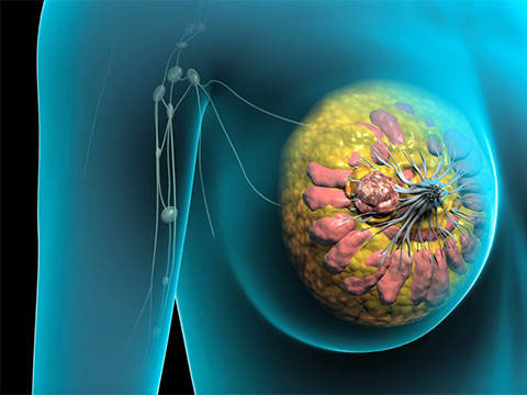
Ultrasound lags behind MRI for supplemental breast cancer screening

Key points
- Cancer screening of women with dense breast tissue is a subject of great interest to both the medical community and the press.
- Breast magnetic resonance imaging (MRI) uses magnetic fields to create an image of the breast.
- A breastultrasound uses sound waves to make a picture of the tissues inside the breast.
- Although supplemental screening via ultrasound is unaffected by breast density, is not associated with ionizing radiation, and does not require IV contrast material, acceptance of this modality has lagged.
Cancer screening of women with dense breast tissue is a subject of great interest to both the medical community and the press. Dense parenchyma reduces the sensitivity of mammography to half that of fatty breasts. Approximately 40% of women 40 years of age or older have dense breast tissue, making supplemental breast cancer screening essential.
Although supplemental screening via ultrasound is unaffected by breast density, is not associated with ionizing radiation, and does not require IV contrast material, acceptance of this modality has lagged.
According to Ellen B. Mendelson, professor of radiology at Northwestern University Feinberg School of Medicine, and Wendie A. Berg, professor of radiology at Magee-Womens Hospital of UPMC, a significant factor is lack of available intensive training opportunities.
"The most common alternative screening modality, MRI, cannot be used with women who have pacemakers or other devices, severe claustrophobia, or renal insufficiency," say Drs. Mendelson and Berg.
"To realize ultrasound's potential to increase the number of cancers detected, intensive training programs need to be put in place for physician performers and interpreters for both handheld and automated breast ultrasound systems."
Regular breast cancer screening is important for all women, but even more so for those at higher risk. If you are at higher risk of breast cancer, you may need to be screened earlier and more often than women at average risk.
A woman is considered at higher risk if she has one factor that greatly increases risk or several factors that together increase risk. Your health care provider may use different tools to assess your risk and help you make a personalized breast cancer screening plan.
What is breast MRI?
Breast magnetic resonance imaging (MRI) uses magnetic fields to create an image of the breast. Breast MRI is more invasive than mammography because a contrast agent is given through an IV before the test.
Breast MRI and breast cancer screening
At this time, breast MRI is mostly used in breast cancer diagnosis and staging, rather than in screening. However, there is growing evidence that breast MRI in combination with mammography is better than mammography alone at finding breast cancer in certain women at higher risk. ACS and NCCN recommend screening with mammography plus breast MRI for some women at higher risk of breast cancer, including those with:
A BRCA1 or BRCA2 gene mutation
A first-degree relative with a BRCA1/2 mutation, but personally have not been tested for BRCA1/2 mutations
A strong family history of breast or ovarian cancer (for example, two or more first-degree relatives with breast cancer or two or more with ovarian cancer) to the chest area before age 30Li-Fraumeni, Cowden or Bannayan-Riley-Ruvalcaba syndrome (or a first-degree relative with one of these syndromes or family with a known p53 or PTEN gene mutation)A greater than 20 percent lifetime risk of invasive breast cancer
Written and revised by
Gerald Hurowitz, M.D.
Our doctors write and review articles for accuracy and keep them up to date.
health.am provides general information, not medical advice. Always consult a qualified health professional about your own health.
Explore health topics
More in this topic
Cancer → News
Nonrecommended screenings for prostate, breast cancer in older individuals
An estimated 15.7 percent of individuals 65 or older may have received nonrecommended screenings for prostate and breast cancers because they had limited life expectancies of less than 10 years, according to an article…
22 January 2016
 Guide
New method for better treatment of breast cancer
A new study shows that a novel imaging-based method for defining appropriateness of breast cancer treatment is as accurate as the current standard-of-care and could reduce the need for invasive tissue sampling. The…
31 December 2015
 News
T cells that recognize HER2 teceptor may prevent HER2+ breast cancer recurrence
Recurrence of HER2-positive breast cancer after treatment may be due to a specific and possibly cancer-induced weakness in the patient's immune system - a weakness that in principle could be corrected with a…