Lymphoepithelial Cyst of the Mediastinum: study

Key points
- We report a rare case of lymphoepithelial cyst of the mediastinum.
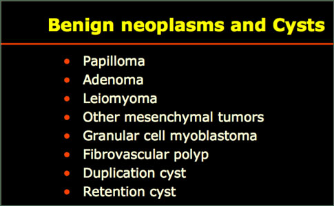
- Benign neoplastic cysts are generally congenital and account for approximately 20% of primary mediastinal lesions, the most common of which are bronchogenic cysts (50%–60%), followed by pleuropericardial cysts (20%–30%), and then by enteric or duplication cysts (7%–15%).
- Ishimaru Y, Shibata Y, Ohkawara S, Ohshima H, Kihara S.
- Histopathologic examination showed that the cystic mass was lined with 1 layer of ciliate columnar epithelium.
We report a rare case of lymphoepithelial cyst of the mediastinum. A 38-year-old woman was found to have a right paratracheal mediastinal mass on chest radiograph. Computed tomographic scanning showed a cystic mass on the right side of the trachea. The encapsulated mass was situated in the upper mediastinum and was adherent to the trachea.
The lesion was resected via a right thoracotomy. Histopathologic examination showed that the cystic mass was lined with 1 layer of ciliate columnar epithelium. There were no malignant foci. These findings were consistent with a diagnosis of multicystic lymphoepithelial cyst.
The postoperative course was uneventful, and the patient was discharged from the hospital on the 7th postoperative day.
Primary cysts of the mediastinum include bronchogenic cysts, enteric or duplication cysts, mesothelial cysts, thymic cysts, and thoracic duct cysts. Mediastinal cystic lesions are generally congenital in origin and form around the 6th week of gestation, due to abnormal development of the tracheobronchial tree, the primitive intestine, or both. To the best of our knowledge, there are only 2 other reports of lymphoepithelial cyst of the mediastinum in the English-language medical literature. We present the case of a 38-year-old woman who had a lymphoepithelial cyst in the mediastinum, which was detected by means of routine chest radiography.
In April 2004, a 38-year-old asymptomatic woman underwent routine chest radiography, which revealed a mass in the upper mediastinum. Computed tomographic (CT) scanning of the chest revealed a well-circumscribed multicystic lesion, about 5.0 cm in diameter, in the right upper paratracheal region. The results of serum tumor marker investigations were as follows: CA-125 antigen, 103.5 U/mL; and β-HCG (human chorionic gonadotropin), 4 mIU/mL. Other laboratory and examination results were within normal limits.
A right thoracotomy was performed. A multicystic, capsulated mass was found in the right upper mediastinum. The dimensions of the cystic mass were 4 × 5 × 6 cm3. The cystic mass, which did not contain fluid, was adherent to the trachea, with no communication to it. The mass was totally excised. On microscopic examination, the mass was seen to consist of microcysts that were lined by a ciliated columnar epithelium and surrounded by lymphoid tissue with germinal centers. Upon immunohistochemical testing, the cyst's epithelioid cells were strongly positive for cytokeratin. The cystic mass was diagnosed as a lymphoepithelial mediastinal cyst.
The postoperative course was uneventful, and the patient was discharged from the hospital on the 7th postoperative day. Follow-up was performed at the 3rd, 6th, and 12th postoperative months, and no radiologic change was observed.
Discussion
Benign neoplastic cysts are generally congenital and account for approximately 20% of primary mediastinal lesions, the most common of which are bronchogenic cysts (50%–60%), followed by pleuropericardial cysts (20%–30%), and then by enteric or duplication cysts (7%–15%). To our knowledge, this is the 3rd case described in the English medical literature. The 1st case was that of a lymphoepithelial cystic lesion related to adenocarcinoma. The 2nd case was that of a lymphoepithelial cyst in the mediastinum with an opening to the trachea. In our case, the patient had a lymphoepithelial cyst in the right upper mediastinum and bronchiectasis of the left lower lobe. The cysts previously described in the literature may have derived from a remnant of the 4th bronchial pouch. A tentative diagnosis of bronchogenic cyst is often made by considering the location of the lesion, because a bronchogenic cyst would be found in the upper compartment of the mediastinum. However, extension into an adjacent area is common, and the final diagnosis usually requires biopsy or resection.
Written and revised by
Andrey Nailor, M.D.
Our doctors write and review articles for accuracy and keep them up to date.
health.am provides general information, not medical advice. Always consult a qualified health professional about your own health.
Explore health topics
More in this topic
Cancer → News
Certain types of polyps may warrant keeping closer tabs on the colon
Being on the lookout for certain features of polyps may help physicians keep a closer eye on patients at risk for colorectal cancer. Starting at age 50, or earlier with certain risk factors, patients are advised to be…
15 April 2016
 News
Smartphones could improve skin cancer detection in developing countries
Everyone knows smartphones can be used as calendars, calculators, radios and cameras. But, did you know they can also be used as microscopes that have the potential to save lives? They are called smartphone microscopes…
17 March 2016
 News
Study shows broccoli may offer protection against liver cancer
Consumption of broccoli has increased in the United States over the last few decades as scientists have reported that eating the vegetable three to five times per week can lower the risk of many types of cancer…