Technologies give high-resolution 'snapshot' of cancer tissues

Key points
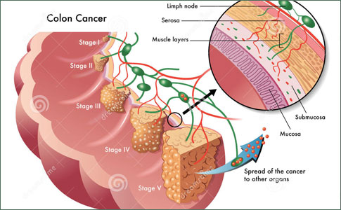
- Stanford researchers have melded tools and technologies from engineering, computer science and stem cell biology to analyze hundreds of individual cancer cells and draw the most accurate portrait yet of the cellular composition of human colon cancer tissues.
- In doing so, they have shown that the development of cancer is a kind of caricature of normal tissue development, and have discovered markers that allow them to gauge more accurately how dangerous a cancer is likely to be.
- In tumor tissues, not all cancer cells are created equal.
- “We have spent the last several years developing single-cell genomic measurement technologies,” said Quake, the Lee Otterson Professor in the School of Engineering and a professor of bioengineering and of applied physics.
Stanford researchers have melded tools and technologies from engineering, computer science and stem cell biology to analyze hundreds of individual cancer cells and draw the most accurate portrait yet of the cellular composition of human colon cancer tissues.
In doing so, they have shown that the development of cancer is a kind of caricature of normal tissue development, and have discovered markers that allow them to gauge more accurately how dangerous a cancer is likely to be. They hope the work will lead to better and more targeted cancer therapies.
In tumor tissues, not all cancer cells are created equal. Solid tumors usually contain many kinds of cancer cells, each of which might be more or less dangerous, and may vary in how they respond to various anti-cancer therapies. One major problem researchers have had in understanding cancer is that when they analyze a cancerous lesion, they usually look at whole-tumor samples that contain a mixture of all these cells types. Amidst this diversity, spotting the signs that betray the really dangerous cells is difficult, like trying to draw the psychological profile of a murderer by sending out questionnaires to everyone who ever committed a legal infraction.
In a paper published online Nov. 13 in Nature Biotechnology, a team led by Stephen Quake, PhD, and Michael Clarke, MD describes how they used the single-cell PCR microfluidic technology invented in the Quake laboratory to analyze the individual gene-expression profile of hundreds of single colon cancer cells, which then they grouped into different subtypes.
“We have spent the last several years developing single-cell genomic measurement technologies,” said Quake, the Lee Otterson Professor in the School of Engineering and a professor of bioengineering and of applied physics. “To finally get all the tools together and produce a biologically important result is very gratifying.”
Using these technologies to analyze the gene-expression patterns of single cells, one by one, allows the researchers to dissect their different identities and clarify the physiology of complex tissue systems, explained Piero Dalerba, MD, instructor of medicine and co-first author of the study. The two other first authors are research associate Tomer Kalisky, PhD, and Debashis Sahoo, PhD, instructor of pathology. Taking advantage of a computer search technique newly developed by Sahoo, the authors identified 47 differentially expressed genes that are useful for the visualization of the various kinds of cells in the tumor.
“In a single experiment, we can now take a ‘snapshot’ of the cell composition of a specific tissue, visualize its different cellular subsets and easily discover novel markers to define them,” said Kalisky.
This methodology is important for the study of minority populations of cells, such as stem and progenitor cells, said Clarke, professor of oncology and the Karel H. and Avice N. Beekhuis Professor in Cancer Biology. “Using these techniques, we have cut a decade off the attempt to understand cellular hierarchy in tissues.”
Written and revised by
Andrey Nailor, M.D.
Our doctors write and review articles for accuracy and keep them up to date.
health.am provides general information, not medical advice. Always consult a qualified health professional about your own health.
Explore health topics
More in this topic
Cancer → News
High cholesterol fuels the growth and spread of breast cancer
A byproduct of cholesterol functions like the hormone estrogen to fuel the growth and spread of the most common types of breast cancers, researchers at the Duke Cancer Institute report. The researchers also found that…
29 November 2013
 News
High-fat diet during puberty speeds up breast cancer development
New findings show that eating a high-fat diet beginning at puberty speeds up the development of breast cancer and may actually increase the risk of cancer similar to a type often found in younger adult women. The…
27 November 2013
 News
Higher than normal levels of Vitamin B12 may indicate cancer risk
Vitamin B12 (cobalamin [Cbl]) is essential for maintaining healthy bodily function but higher than normal levels (reference range 200-600 pmol/L) may indicate that a patient is at risk of developing certain cancers,…