David Cohen, M.D.·Published 30 May 2008·15 min read
Key points
Gene therapy for advanced breast cancer is anticipated to be a useful therapeutic approach.
The carcinogenic process requires an accumulation of multiple gene mutations or abnormalities of gene expression.
Breast cancer cells have long been supposed to have low antigenecity and to be resistant to immune therapy.
Transfer of drug-activating enzyme gene into tumor cells and treatment with a prodrug form of chemotherapeutic agents causes a high concentration of the activated drug in the tumor tissue and apoptosis of tumor cells.
Review of Clinical Gene Therapy Trials for Breast Cancer and MDR1 Gene Therapy Trial in Cancer Institute Hospital
Gene therapy for advanced breast cancer is anticipated to be a useful therapeutic approach. Strategies in ongoing clinical protocols can be divided into four groups: (1) suppression of oncogenes or transfer of tumor-suppressor genes; (2) enhancement of immunological response; (3) transfer of suicide genes; (4) protection of bone marrow using drug resistance genes. We have started a clinical study of multidrug resistance (MDR1) gene therapy. Advanced breast cancer patients received high dose chemotherapy and autologous peripheral blood stem cell transplantation (PBSCT) with MDR1-transduced hematopoietic cells, and then were treated with docetaxel. Two patients have been treated so far, and in vivo enrichment of MDR1-transduced cells with docetaxel treatment has been seen. Both patients are in complete remission and had no apparent adverse effects from the MDR1 gene transfer. Breast Cancer 13:8-15, 2006.
The cure rate of advanced or recurring breast cancer is under 5%, so the usual goal of treatment is prolongation of survival or improvement of quality of life (QOL), not cure1). Endocrine therapy for hormone-receptor-positive patients, chemotherapy, radiation therapy, bisphosphonates for bone diseases, and trastuzumab for HER2-overex-pressed patients, have all been shown to be effective for advanced breast cancer, but none has been shown to increase the cure rate.
Gene therapy for advanced breast cancer is expected to be a useful therapeutic approach.
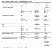
Strategies in ongoing clinical protocols can be divided into four groups: (1) suppression of oncogenes or transfer of tumor-suppressor genes; (2) enhancement of immunological response; (3) transfer of suicide genes; (4) protection of bone marrow using drug resistance genes (Table 1)2, 3).
There are three major methods for gene transfer: (1) transduction of naked DNA such as lipofection (transient expression); (2) transduction of adenoviral vector or vaccinia virus vector (transient expression); (3) transduction of retroviral vector (stable expression). In this paper, ongoing clinical trials of gene therapy for breast cancer are reviewed, and a clinical trial of multiple drug resistance 1 (MDR1) gene therapy at our institution is described.
Present Status of Clinical Trials of Gene Therapy for Breast Cancer
Written and revised by
David Cohen, M.D.
Our doctors write and review articles for accuracy and keep them up to date.
health.am provides general information, not medical advice. Always consult a qualified health professional about your own health.
Suppression of Oncogene Expression or Transfer of Tumor-Suppressor Gene
The carcinogenic process requires an accumulation of multiple gene mutations or abnormalities of gene expression. Common gene abnormalities in breast cancer include p53 gene mutation, ErbB2/ HER2 gene amplification, c-myc gene amplification, and cyclin D1 gene amplification4). Several clinical trials aim to improve those gene abnormalities by local or systemic gene transfer.
A) Transfer of the normal p53 gene: Mutations of the p53 gene are the most frequently found gene abnormalities among various malignancies, including breast cancer5).
Table 1. Clinical Studies of Gene Therapy for Breast Cancer. Tumor cells with mutated p53 genes show defects of cell-cycle regulation, and transfer of normal p53 genes causes cell-cycle arrest or apoptosis. Clinical studies of p53 gene therapy using adenoviral vectors (Advexin, Introgen et al.) for various tumor types, including breast cancer, are ongoing. Von Mehren and Cristo-fanilli have begun clinical studies of a combination of local injection of p53-adenoviral vector into skin metastatic lesions or locally advanced breast cancer and systemic chemotherapy. Baynes has initiated a clinical study of high dose chemotherapy associated with transplantation of autologous peripheral blood stem cells (PBSC) that have been purged ex vivo by p53-adrnovirus infection. Baynes's group has shown that p53 gene transfer has no effect on normal PBSC.
B) Suppression of the ErbB2/HER2 gene: The ErbB2/HER2 gene encodes an 185 kD protein and is a member of the epidermal growth-factor receptor family. This gene is amplified in 20-30% of breast cancer patients, and correlates with a poor prognosis and resistance to hormone therapy4).
Monoclonal humanized murine antibody to ErbB2/HER2 protein (trastuzumab/Herceptin™) is effective in advanced, ErbB2/HER2-overexpressing breast cancer patients6). The adenovirus type 2 or type 5 E1A gene inhibits expression of the ErbB2/HER2 gene, and E1A gene transfer into ErbB2/ HER2-overexpressed tumors causes tumor reduction and enhances sensitivity to chemotherapy in vitro and in vivo7). At MD Anderson Cancer Center, patients with breast cancer or ovarian cancer overexpressing ErbB2/HER2 were treated with gene therapy using a local injection of E1A gene-liposome into skin lesions or pleural/peritoneal effusion8). There was no serious adverse effect other than fever or pain at the injection sites. In six cases in which tumor cells in body fluids could be analyzed, reduction of ErbB2/HER2 expression and a decrease in tumor cells were shown.
E1A gene transfer also reduced tumor growth of non-HER2-overexpressing cells, and E1A gene transfer to tumor tissues of breast cancer or head and neck cancer by lipofection showed minor response in HER2-negative tumors9).
C) Suppression of c-myc and c-fos gene: Arteaga and Holt made a retroviral vector which over-expresses antisense mRNA to c-myc and c-fos genes under the control of mammary tumor virus (MMTV) promoter. Transfer of this vector into a breast cancer cell line suppressed tumor formation in animal models10). They have started a clinical trial of gene therapy for malignant effusion or meningitis in breast cancer patients who have failed standard therapy. Effusions will be drained and replaced with a solution of the vector, then periodically drained to follow the disease and assess gene transfer11).
D) Transfer of melanoma differentiation associated protein 7 (MDA-7): MDA-7 is a novel tumor suppressor gene, and its transfer into tumor cells causes growth suppression and apoptosis. However, MDA-7 gene transfer into normal cell lines does not12). A clinical trial of gene therapy that injects MDA-7- adenoviral vector (Ad-mda7, ISGN 241) into tumor cells has started (Buchholz).
There was no serious adverse effect in a phase I study, and a combination phase I/II study with irradiation has begun.
{pagebreak}
Augmentation of Immunological Response to Cancer Cells
Breast cancer cells have long been supposed to have low antigenecity and to be resistant to immune therapy. So far, reports of nonspecific immune therapies such as BCG have shown that those therapies are not effective for breast cancer13). But since the 1990s, many breast cancer-associated antigens have been reported, and various clinical studies of specific immune therapy for breast cancer, such as vaccination therapy targeted to ErbB2/HER2, are ongoing14, 15). Immune therapy by gene transfer includes: 1) transfer of cytokine genes that enhance immune response, 2) transfer of co-stimulatory molecule genes, and 3) transfer of antigen molecule genes.
A) Transfer of cytokine genes
i) Interleukin-2 (IL-2): Injection of IL-2 gene-adenoviral vector into tumor tissues16), or subcutaneous injection of inactivated tumor cells that were transduced ex vivo by IL-2 gene lipofection (Lyerly) may cause a systemic immune reaction in tumor cells. In a phase I/II study, Stewart et al.17) treated 23 cases with breast cancer or malignant melanoma by injection of 10 7-1010pfu adenovirus-IL-2 into subcutaneous tumors. There was no side effect other than local inflammation of injection sites, and reduction in diameter of subcutaneous tumors was reported in 24% of patients, but there was no PR.
ii) Interleukin-12 (IL-12): Retroviral transfer of IL-12 gene into skin fibroblasts of patients ex vivo, then injection of the fibroblasts into tumor tissues may activate a tumor-specific immune response.
In a phase I study, nine cases with advanced neoplasm including breast cancer were treated by Kang et al. Reduction of tumor at injection sites was shown in four cases, and reduction of tumor at remote sites was shown in one melanoma case.
There was no side effect other than slight pain at the injection sites18).
iii) Granulocyte-macrophage colony stimulating factor (GM-CSF): Retroviral transfer of GM- CSF gene into tumor cells and injection of those cells into subcutaneous tissue may activate systemic immune reaction to tumor cells (Suzuki). The same gene therapy for renal cell cancer has been done in Japan.
iv) Tumor necrosis factor (TNF): Retroviral transfer of TNF gene and Neo gene into tumor cells ex vivo and subcutaneous injection of tumor cells may activate systemic immune response to tumor cells19).
B) Transfer of co-stimulatory molecule gene: Transfer of T cell co-stimulatory molecule CD80 (B7.1) gene into tumor cells by lipofection and injection of those tumor cells into subcutaneous tissue (Urba), or direct injection of CD80-adenoviral vector into tumor tissue (Schuchter) may activate T cell growth and immune response.
C) Transfer of antigen gene: Clinical studies of MUC1(CA15-3) gene transfer by vaccinia virus into tumor cells and injection of tumor cells into subcutaneous tissue (Kufe), simultaneous transfer of MUC1 and CD80 gene (Eder), or HER2 gene transfer (Patel), have been ongoing. Scholl et al. repeatedly administered vaccinia virus containing MUC1 and IL-2 genes (TG1031) intramuscularly to patients with metastatic breast cancer. In 31 patients, two patients (6%) had PR and 15 patients had SD20).
{pagebreak}
Suicide Gene Therapy
Transfer of drug-activating enzyme gene into tumor cells and treatment with a prodrug form of chemotherapeutic agents causes a high concentration of the activated drug in the tumor tissue and apoptosis of tumor cells. Not only transduced cells, but also circumferential cells are reported to die with this gene therapy (bystander effect).
A clinical trial of retroviral herpes simplex virus thymidine kinase (HSV-TK) gene transfer into breast cancer tumor tissues and treatment with gancylovir is ongoing (Favrot).
A phase I study of injection of HER2 promoter-driven cytosine deaminase (CD) gene plasmid into metastatic skin lesions of breast cancer and treatment with prodrug (fluorocytosine) has been reported. Fluorocytosine is transformed into 5FU by the CD gene. Expression of the CD gene in HER2-positive tumor cells has been shown in 9/11 cases at day 2 and 3/10 cases at day 7. Tumor reduction was shown in 4 of 12 cases21).
Retroviral P450 2B6 (CYP2B6) gene transfer into metastatic cutaneous tissues and oral cyclophosphamide therapy causes efficient conversion of prodrug cyclophosphamide into active metabolite phosphoramide mustard in the tumor tissues.
In a phase I study, nine breast cancer and three melanoma patients were treated with CYP2B6 vector (MetXia-P450). One breast cancer patient had a PR and four (33%) had stable diseases (SD) ≥ 3 months22).
{pagebreak}
Bone Marrow Protection by Drug-Resistance Gene
Breast cancer is sensitive to chemotherapy. Response rates of advanced breast cancer for most combination chemotherapy are between 40% and 70% (complete response (CR) rate 10-30%), but duration of response is 7-10 months for PR, and 9-18 months for CR. High dose chemotherapy with autologous blood stem cell transplantation for advanced breast cancer has shown high complete response rates (up to 50%), and 10-15% patients have enjoyed durable remission23, 24). However, most patients will relapse after transplantation.
Randomized studies comparing high dose chemotherapy and conventional chemotherapy showed that median survival times appear to be no better than those achieved with conventional chemotherapy, so far25). Probably high dose chemotherapy cannot completely eradicate residual disease, and insufficient bone marrow function after the reconstitution is a major problem in post-transplantation chemotherapy. One approach to overcome the current situation would be the transplantation of the drug-resistant gene-transduced hematopoietic stem cells so that normal bone-marrow cells will be protected from the toxic effect of anticancer drugs.
A multidrug resistance 1 (MDR1) gene was cloned from cancer cell lines resistant to various anticancer drugs26). The MDR1 gene product (P- glycoprotein, P-gp) is a 170 kD glycoprotein consisting of two trans-membranous domains and two ATP-binding domains. P-gp ATP-dependently excretes various drugs such as doxorubicin, vinka-alkaloids, or taxanes from cytoplasm to extra-cellular fluid. Ex vivo transfer of MDR1 genes into hematopoietic stem cells and transplantation might make post-transplant chemotherapy feasible.
Chemotherapeutic drugs such as docetaxel and paclitaxel, which have good clinical activity in the treatment of breast cancer and are efficiently effluxed by P-gp, might be the best choice for this strategy. Using a retroviral vector, Sorrentino et al.27) transplanted MDR1-transduced bone marrow into irradiated mice and then treated them with paclitaxel. Paclitaxel treatment increased MDR1- transduced leukocytes in peripheral blood (in vivo amplification), and MDR1-transduced mice showed reduced bone marrow suppression by paclitaxel (bone marrow protection). Then, several groups have undertaken clinical studies of MDR1 gene therapy for advanced breast cancer or other neoplasms28-30).
A group at MD Anderson Cancer Center first reported the results of clinical trials28). They performed retroviral gene transfer without using cytokines, and in suspension or with autologous stromal cells. In vitro transduction efficiency was 2.8% with the solution method and 5.6% with the stromal method, detected by in situ PCR. But three to four weeks after transplantation, direct PCR assay of peripheral blood leukocytes in patients showed positive results in 0/10 with the solution method, and 5/8 with the stromal method.
These data show insufficient transduction efficiency without using cytokines. NCI also reported the results of a clinical trial of retroviral MDR1 gene therapy30). They transferred MDR1 genes into bone marrow mononuclear cells or peripheral blood stem cells stimulated by IL-3, IL-6, and SCF. Ex vivo transduction efficiency was 0.2-0.5%.
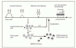
Fig 1. Schema of MDR1 gene therapy for advanced breast cancer patients in Cancer Institute Hospital. They treated transplanted patients with paclitaxel, but they could not show any enrichment of MDR1- transduced white blood cells by PCR. A group at Columbia University also transferred MDR1 genes into bone marrow mononuclear cells or peripheral blood stem cells stimulated by IL-3, IL-6, and stem cell factor (SCF). They showed that 20-70% of BFU- E or CFU-GM colonies from transferred CD34- positive cells were positive for MDR1 by PCR. BM from patients 3-12 weeks after transplantation showed MDR1-positivity by PCR in 2/5 patients.
They also analyzed P-gp expression in bone marrow cells using flow cytometry, but they could not show any expression. Clinical studies of MDR1 gene therapy are now ongoing at several institutions (Stewart, Cowan, Disseroth, Hesdorffer, O'Shaughnessy).
{pagebreak}
MDR1 Gene Therapy in Cancer Institute Hospital
Our group also started MDR1 gene therapy for breast cancer. This study was approved by the Ministry of Health and the Ministry of Education and Science on February 24, 2000. The outline of the protocol is shown in Fig 1. We selected histologically confirmed, metastatic breast cancer patients who achieved good PR or CR to a precedent conventional dose chemotherapy regimen (using anthracycline and/or taxane). We used a HaMDR vector in which wild type MDR1 cDNA (Kyoto University) had been inserted into pHa vector (NCI) derived from Harvey mice sarcoma virus (HaMSV). Peripheral blood stem cells (PBSC) were harvested by cyclophosphamide and G-CSF.
CD34-positive cells were selected from about one third of PBSC, and HaMDR was transferred into those cells stimulated by SCF, thrombopoietin, IL-6, Flt-3 ligand, and soluble IL-6 receptor. Transduced PBSC were checked for safety (presence of replication-competent retrovirus, etc.) and then frozen. Patients were treated with high-dose cyclophosphamide, thiotepa, and carboplatin. Then unprocessed and MDR1 gene-transduced PBSC were transplanted together. After bone marrow was reconstituted and patient status was normalized, patients were treated with 50% of standard dose docetaxel, then with increased doses up to 100% if grade 4 neutropenia was not recorded .
Gene transfer efficiency and P-gp expression were checked with PCR and flowcytometry analysis, using peripheral leukocytes and bone marrow cells.
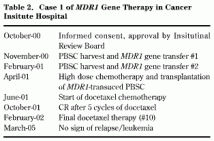
So far, two patients have finished high-dose chemotherapy, PBSC transplantation with MDR1 gene transfer, and then docetaxel chemotherapy (Table 2).
Table 2.Case 1 of MDR1 Gene Therapy in Cancer Insitute Hospital Peripheral blood P-gp-positive leukocytes increased to 5% after transplantation but decreased gradually. During docetaxel chemotherapy after transplantation, in vivo expansion of the MDR1-transduced cells (up to 10%) was obser ved. Comparison of two patients suggests the presence of a bone-marrow protection effect by MDR1 expression during docetaxel chemotherapy, but this is not clear. No serious side effect was observed, and the patients have been in complete remission for 3 years.
Retroviral gene therapy causes random insertion of exogenous genes into genome DNA of target cells, so it may cause carcinogenesis by activation of oncogene or inactivation of tumor suppressor gene. At the end of 2002, occurrence of T cell leukemia in two patients after gene therapy for X- linked severe combined immune deficiency (X- SCID) was reported. A genetic defect in the ϒC gene, which is a common domain of multiple interleukin receptors (IL-2R, IL-4R, IL-7R, et al.), causes severe defects of T cell and natural killer cells as well as severe immune deficiency in X- SCID patients. Retroviral ϒC gene transfer using autologous CD34-positive hematopoietic cells in X- SCID patients restored immune system in 9 of 11 patients31). But T cell leukemia occurred in three patients (one more patient in January 2005) of those 9. In the leukemic cells, retroviral vector was inserted in the LMO2 gene, which causes T cell leukemia32). Then the FDA recommended suspension of all clinical trials of retroviral gene therapy for hematopoietic stem cells. We also suspended MDR1 gene therapy for the third patient in January 2003. After thorough investigation of retroviral gene therapy trials for hematopoietic stem cells all over the world, no leukemia event has been found in clinical gene therapy trials, other than the French X-SCID trial (American Society for Gene Therapy Annual Meeting, 2003). Screening of the Mouse Retroviral Cancer Gene database showed that retroviral insertion into ϒC and LMO2 gene was found in two cases each, and insertion into both genes were found in one case.
This fact suggests that both genes are oncogenes, and that the two genes can collaborate33). In X- SCID gene therapy, a double hit with retroviral activation of LMO2 gene and exogenous activated ϒC gene might be necessary for leukemogenesis.
If so, retroviral gene therapy with non-oncogenic genes might have a low risk of cancer34).
Thereafter, gene therapy using retroviral vector resumed, and retroviral gene transfer into hematopoietic cells of adenosine deaminase deficiency patients was begun in Japan at the end of 2003. We also resumed our MDR1 gene therapy after changing the protocol (informed consent with regard to the adverse effects and more thorough investigation of patients' peripheral blood), and started high-dose chemotherapy and transplantation of PBSC with MDR1 gene transfer to the third patient in July 2004.
We also started investigation of insertion sites of HaMDR vector in the first two patients. A clonality study of leukocytes from case 1 showed eight long-lived clones of MDR1-tranduced hematopoietic stem cells. No sign of expansion of any clones has been observed.
To summarize the data of our own and other institutions' clinical studies of retroviral MDR1 gene therapy, first, there has been no serious side effect, including secondary neoplasm, but thorough investigations including retroviral insertion sites are necessary. Second, maintenance of MDR1-transduced hematopoietic cells for more than one year was confirmed. Third, the MDR1- transduced cells were selectively enriched in vivo by chemotherapy. Whether MDR1 gene therapy can protect bone marrow from chemotherapy is not yet certain. We have almost finished proof-of-concept stage for the gene therapy, and we should be able to show clinical benefits compared with conventional therapy.
The techniques and knowledge of gene therapy are still limited, so we must proceed with caution, and we must inform patients of both the risks and benefits of the therapy.
Acknowledgements
This work was supported by the grants from the Ministry of Health, Labor and Welfare, and the Ministry of Education, Culture, Sports, Science and Technology and the Nippon Foundation, Japan.
Shunji Takahashi1, Yoshinori Ito1, Kiyohiko Hatake1, and Yoshikazu Sugimoto2,3
1 Department of Medical Oncology, Cancer Institute Hospital and 2 Department of Gene Therapy, Cancer Chemotherapy Center, Japanese Foundation for Cancer Research, 3 Department of Chemotherapy, Kyoritsu University of Pharmacy, Japan.

 News
 News
 News